Figures & data
Table 1. List of putative rickettsial virulence genes.
Figure 1. Rickettsial intracellular lifecycle.
1. Rickettsia attaches to host cells via interaction of rickettsial surface proteins with host cell receptors; 2. Downstream signaling cascades lead to the internalization of Rickettsia; 3. Using several membranolytic factors, Rickettsia escapes the endocytic vacuoles; 4. Rickettsia resides within the host cytosol and replicates while taking nutrients from the host; 5. Some Rickettsia species exhibit actin-based motility; 6. Rickettsia undergoes cell-to-cell spread; 7. Rickettsia breaks double vacuolar membrane structures; 8. Rickettsia resides within the cytosolic compartment and reinitiates the infectious cycle.
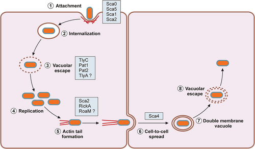
Figure 2. Secretion systems in Rickettsia.
Rickettsia utilizes five secretion pathways: Sec-T5SS, Sec-TolC, Tat, T1SS, and T4SS. Most Sca proteins have three domains: the N-terminal secretion signal (black), passenger domain (blue), and C-terminal autotransporter domain (red). The black and purple boxes on RARP-1 denote the N-terminal secretion signal and C-terminal Ankyrin repeat domain. The C-terminal Ankyrin repeat domain of RARP-2 is colored in yellow. Question marks 1 and 2 indicate uncharacterized proteins that travel through the Tat and/or T1SS. Question mark 3 indicates divergent destinations of RARP-1 in Rickettsia. Lastly, question mark 4 indicates unresolved compositions of S-layer proteins and their interactions with the outer membrane components, such as O-Ag. IM-inner membrane; PP-periplasm; PG-peptidoglycan; OM-outer membrane; LPS-lipopolysaccharide; S-layer-surface layer.
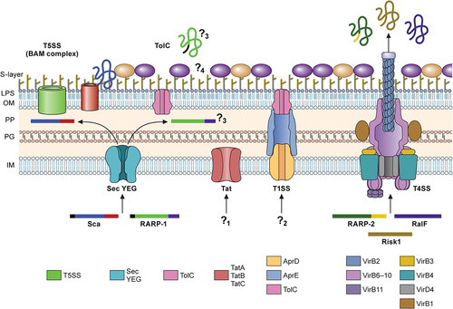
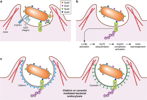
Figure 3. Rickettsial determinants in host cell attachment and internalization.
A. Rickettsial entry into mammalian cells is mediated by interactions of its surface antigens- Sca0 with α2β1 integrin and FGFR1 receptor, Sca5 with Ku70, and Sca1 and Sca2 with unknown receptors on the cell surface; B. Ku70-Sca5 interaction leads to activation of E3 ubiquitin ligase c-Cbl causing ubiquitination of Ku70. This initiates several downstream host-signaling pathways leading to activation of the Arp2/3 complexes resulting in actin rearrangement; C. This induces bacterial internalization via actin, clathrin, and caveolin 2- mediated endocytosis.