
Figures & data
Figure 1. Role of OmpR in A. baumannii virulence assessed in thigh and septicaemia mouse infection models.
A) and B) Neutropenic CD-1 male mice (12 per group) were infected using thigh injection of 107 cfu of AB5075 wildtype (black) or AB5075 ΔompR mutant (white) and 4 animals per group were sacrificed at 2, 24 and 48 hours post infection for bacterial titers determination in A) thigh and B) blood. Mortality was observed in the wildtype group at 24 and 48 hours enabling bacterial titers analysis on only 3 and 1 animal, respectively. Unpaired t-test, *P-value < 0.05. C) Immunocompetent C57BL6J male mice (10 per group) were infected with intravenous injection of 5 × 10⁷ cfu of HUMC1 wildtype (black) or HUMC1 ΔompR mutant (white) and mice survival was monitored for 7 days. Log-rank (Mantel-Cox) test, *P-value < 0.05.
Figure 2. Role of OmpR in A. baumannii virulence assessed in G. mellonella infection model.
A) Groups of 10 larvae were infected with 105 cfu of AB5075, HUMC1, AYE or BV191 wildtype strains (blue) and their isogenic ΔompR mutants (red). B) Groups of 10 larvae were infected with 104 cfu of AB5075 wildtype (light blue), AB5075 ΔompR (red) and OmpR complemented (OmpRc, dark blue) strains. C) Groups of 10 larvae were infected with 104 cfu of AB5075 wildtype (light blue), AB5075 ΔompR (red), AB5075 OmpR D71E (green), AB5075 OmpR D71A (orange) and AB5075 OmpR R198L (purple) mutants. Larvae survival was monitored over 72 hours. Log-rank (Mantel-Cox) test compared to wildtype, *P-value < 0.05.
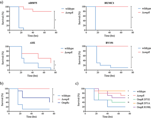
Figure 3. Identification and confirmation of OmpR-regulated genes.
A) RNA-seq results representation of differentially expressed genes between AB5075 wildtype and its ΔompR mutants. Sixty-five OmpR-activated candidate genes (<-2 log2 fold change, blue zone) and 71 OmpR-repressed candidate genes (>2 log2 fold change, green zone) were identified. B) The RNA-seq results were combined with a Tn-seq dataset conducted to identify A. baumannii genes that are important or deleterious for A. baumannii virulence [Citation26]. Nineteen OmpR-activated candidate genes were important for A. baumannii virulence whereas 12 OmpR-repressed candidate genes were deleterious for A. baumannii virulence. RR: read ratio. C) The transcript levels were determined in A. baumannii AB5075 wildtype, AB5075 ΔompR (white), AB5075 OmpR D71E (light grey), AB5075 OmpR D71A (dark grey) and AB5075 OmpR R198L (black) mutants by quantitative real-time PCR and the expression level was normalized to the expression of the AB5075 wildtype strain (means ± SEM of two technical replicates). D) The transcript levels of ABUW_RS02965 (white) and ABUW_RS13110 (black) were determined in A. baumannii ΔompR mutants and normalized to the transcript levels of their respective wildtype strains (means ± SEM of two technical replicates). Horizontal dotted lines depict a 2-fold up- or downregulation.
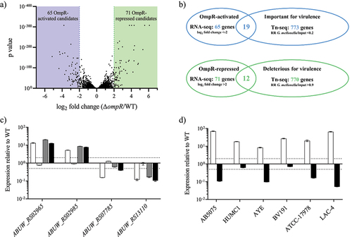
Figure 4. Study of OmpR protein-DNA binding using D-ELISA and in silico analysis.
OmpR binding to the F1aF1b oligonucleotide (2 µg/mL) was assessed with 3.1 µg/mL (light grey), 6.2 µg/mL (dark grey) and 12.5 µg/mL (black) of the OmpRECO (A) and OmpRABA (B) proteins. A negative control without protein was included (white). Unpaired t-test compared to control, *P-value < 0.05. C) The binding of OmpRABA and OmpRABA R198L mutant (6 µg/mL) to the F1aF1b oligonucleotide (2 µg/mL) was tested under unphosphorylated (white) and phosphorylated (black) conditions. Unpaired t-test compared to wildtype, *P-value < 0.05. D) A 10 nucleotide motif (highlighted in red) common to all known OmpRECO DNA binding sites upstream of ompF (F1aF1b, F2aF2b, F3aF3b and F4aF4b) and ompC (C1aC1b, C2aC2b and C3aC3b) was identified in the intergenic region upstream of the 4 OmpRABA regulated genes using the MEME suite [Citation30]. The weblogo of the sequence motif is depicted in the upper right corner. E) OmpRABA binding to the ABUW_RS02965_A, B and C oligonucleotides (2 µg/mL) was assessed with 1.5 µg/mL (light grey), 3 µg/mL (dark grey) and 6 µg/mL (black) of proteins. A negative control without protein was included (white). Only the ABUW_RS02965_A encodes the identified OmpR DNA binding motif (Supplementary Figure S4). Unpaired t-test compared to control, *P-value < 0.05. The data represent the mean ± SD of at least two replicates.
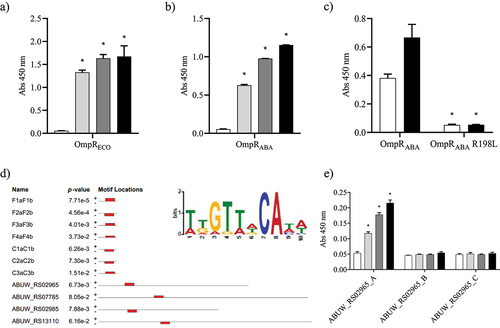
Figure 5. Structure of the DNA-binding domain of A. baumannii OmpR and computational hotspots analysis.
A) Overview of the crystal structure of the DNA-binding domain (DBD) of A. baumannii OmpR. B) Superposed crystal structure of OmpR DBD from A. baumannii (blue) on DNA bound structure of OmpR from E. coli (grey). C) Amino acids (blue colored for OmpRABA and grey colored for OmpRECO) that are involved in making H-bond and salt bridges with DNA are shown. D) The 3D structures of OmpRECO (1opc) and OmpRABA DBD (chain A, B, C and D) are represented and colored based on temperature scale from blue (low ligand interaction probability) to red (high ligand interaction probability). Individual residues with high predicted ligand-binding potential are highlighted. The interaction site is around the conserved residues TVWG(V/L). Hotspot 1 in OmpRABA-DBD chain A contains D218, as anchor residue for ligand binding. Hotspot 2 in OmpRABA-DBD chain D contains residues S222, R223 and R226 (S206, R207 and R210 in OmpRECO DBD). Hotspot 3 in OmpRABA-DBD chain C contains residues K186, L204, and T178/T179.
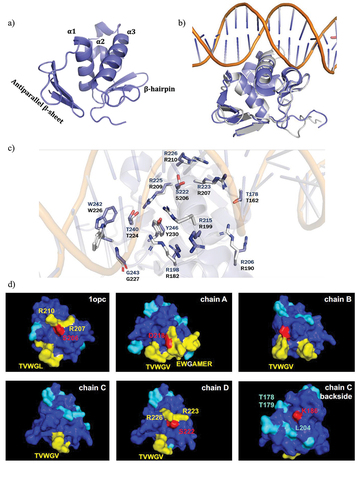
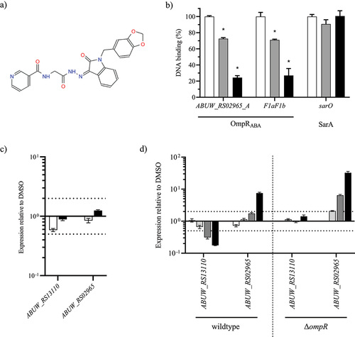
Figure 6. OmpR inhibitory activity of VSIS_039.
A) Structure of the compound VSIS_039 (STL300125, Vitas-M Chemical Limited). B) OmpRABA DNA inhibition was assessed by D-ELISA using 2 µg/mL biotinylated ABUW_RS02965_A and F1aF1b oligos and 6 µg/mL of OmpRABA in the presence of DMSO (white), 10 µM (grey) and 20 µM (black) of VSIS_039 compound (mean ± SD of 2 technical replicates). As negative control, VSIS_039 was also tested on SarA and its cognate DNA binding site using 10 µg/mL of the SarA protein and 2 µg/mL of the biotinylated sar0 oligos. Unpaired t-test compared to DMSO, *P-value < 0.05. C) The transcript levels of ABUW_RS02965 and ABUW_RS13110 were determined in the A. baumannii AYE wildtype strain in the presence of VSIS_039 at 33.3 μM (white) and 100 μM (black). The transcripts levels were normalized to those of the DMSO control (mean ± SEM of 2 technical replicates). D) The transcript levels of ABUW_RS02965 and ABUW_RS13110 were determined in the A. baumannii AYE efflux depleted (ΔadeABC ΔadeFGH ΔadeIJK) wildtype and ΔompR strains in the presence of VSIS_039 at 10 μM (white), 20 μM (light grey), 40 μM (dark grey) and 80 μM (black). The transcripts levels were normalized to those of the DMSO control (mean ± SEM of 2 technical replicates). Horizontal dotted lines depict a 2-fold up- or downregulation.
Supplemental material
Supplemental Material
Download PDF (1 MB)Data availability statement
All data are fully available without restriction.