
Figures & data
Figure 1. Protein structure prediction of pp65 and gH and preparation of coupled peptide vaccines. (a) the coupled diagram of peptide-CRM197. The secondary structure of pp65 (b) and gH (c) were predicted by the online software SOPMA. (d) After coupling the peptides with CRM197, three peptide-CRM197 were subjected to Coomassie brilliant blue staining after SDS-PAGE.
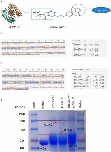
Table 1. Prediction of pp65 epitopes.
Table 2. Prediction of gH epitopes.
Figure 2. Analysis of three peptide-CRM197 vaccines for maturation of DCs in vitro. DC2.4 cells were stimulated with PBS, LPS, and three peptide-CRM197 vaccines for 48 h. The percentages of CD11c+ DCs expressing CD80 (a), CD86 (b), MHCII (c), and CD40 (d) were compared by flow cytometry. At the same time, the cell culture supernatants were harvested to detect the secretion levels of TNF-α (e), IL-6 (f), IL-1β (g), and IL-12p70 (h). All data were expressed as mean ± SD, * means P < .05, ** means P < .01, *** means P < .001, and **** means P < .0001 (one-way ANOVA).
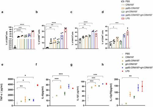
Figure 3. Analysis of three peptide-CRM197 vaccines for maturation of DCs in vivo. The mice were immunized with PBS, inactivated HCMV, and peptide-CRM197 vaccines. On the 3rd day, the expression of CD80, CD86, CD40, and MHCII in CD11c+ cells was analysed by flow cytometry. (a) the histograms of flow cytometric analyses of CD80, CD86, CD40, and MHCII in CD11c+ cells. (b) the percentages of CD11c+ DCs expressing CD80, CD86, CD40, and MHCII. All data were expressed as mean ± SD, * means P < .05, ** means P < .01, *** means P < .001, and **** means P < .0001 (one-way ANOVA).
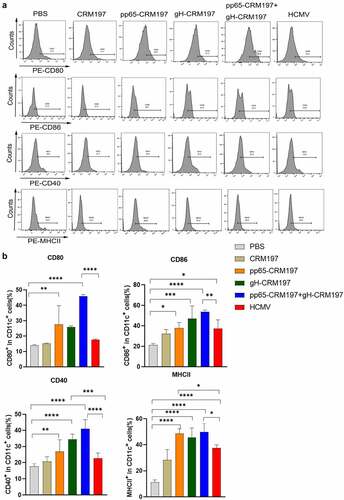
Figure 4. The signalling mechanism of activating DCs by three peptides-CRM197 vaccines. DC2.4 cells were stimulated by PBS, LPS, and peptide-CRM197 vaccines for 48 h, cellular proteins were extracted, and the expression of p-MKK6 (a) and p-p38 (b) were detected by Western blotting. (c) DC 2.4 cells were pretreated with SB203580 for 1 h, then were stimulated with PBS, LPS, and three peptide-CRM197 vaccines for 48 h. The expression of CD80, CD86, MHCII, and CD40 was detected by flow cytometry. All data were expressed as mean ± SD, * means P < .05, ** means P < .01, *** means P < .001, and **** means P < .0001 (one-way ANOVA).
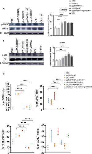
Figure 5. Evaluation of immune responses in three peptides-CRM197 vaccines. On the 35th day after the first immunization, the lymphocyte single-cell suspension was obtained, and the expression of cytokines was analysed by flow cytometry. The MFI expression level of IFN-γ (a), IL-2 (b), and IL-4 (c) in CD3+CD4+T cells, IFN-γ (d), and TNF-α (e) in CD3+CD8+T cells, and Foxp3 (f) in Treg cells. The inactivated HCMV was used to stimulate splenic lymphocytes of mice, and lymphocyte proliferation was detected at 48 and 72 h (g). Detection of neutralizing antibody titre in mice serum (h). All data were expressed as mean ± SD, * means P < .05, ** means P < .01, *** means P < .001, and **** means P < .0001 (one-way ANOVA).
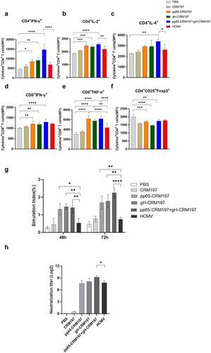
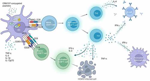
Figure 6. Schematic diagram of the peptide-CRM197 vaccines activating immune responses.
Data availability statement
The authors confirm that the data supporting the findings of this study are available within the article [and/or] its supplementary materials