
Figures & data
Figure 1. The structure and drug resistance development of MPD. (a) The chemical structure of 1-(4-methoxyphenyl)-1hydro-pyrrole-2,5-dione (MPD). (b) Drug resistance development test of MPD and FLC against C. albicans YEM30 using consecutive treatment of 1/2 MIC of MPD or 1/2 MIC of FLC.
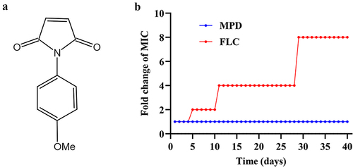
Table 1. Gene-specific primers used for real-time RT-PCR.
Table 2. The MIC values of MPD against different genotypes of C. albicans.
Figure 2. MPD decreases the adhesion of C. albicans. The XTT reduction assay (a) and microscopy (b) were used to detect the inhibitory effect of MPD on the adhesion of C. albicans. The bar in (b) indicates 100 µm. Results in (a) are shown as means ± SDs.
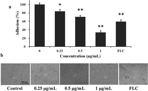
Figure 3. MPD inhibits the hyphal formation of C. albicans. YEM30 strain were cultured in the RPMI 1640 medium and spider solid plate containing different doses of MPD at 37 °C. The imaging was observed by microscope every 3 h. The bar indicates 100 µm.
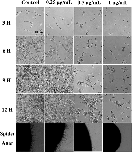
Figure 4. MPD suppresses the biofilm formation of C. albicans. YEM30 strain treated with MPD were incubated in RPMI 1640 medium at 37 °C for 24 h. After incubation, an XTT reduction assay (a), the observation with microscopy and SEM (b) were used to detect biofilm formation. (b) The first row of photographs was taken under the optical microscope and the remaining two rows were taken under the SEM. (c) The effect of MPD on the CSH of C. albicans. The bars in (a), (b), and (c) show 100 µm, 100 μm, and 20 μm, respectively. Results in (a) and (c) are shown as means ± SDs.
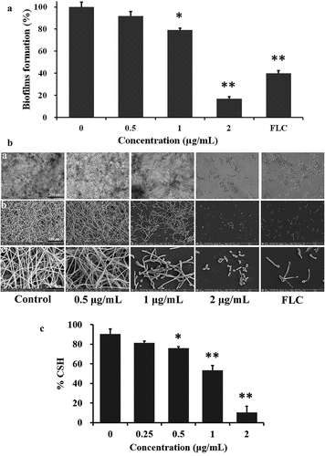
Figure 5. The effect of cAMP on the hyphal and biofilm formation in the presence of MPD treatment. (a) YEM30 strain treated with MPD were incubated in RPMI 1640 medium at 37 °C for 12 h. The intracellular cAMP content was measured by cAMP Elisa kit. (b) YEM30 strain were diluted in RPMI 1640 medium containing different concentrations of MPD, and then added with 1 mM db-Camp. After incubation at 37 °C for 4 h, the imaging was observed by microscopy. The effect of exogenous cAMP on the 1 and 2 μg/mL of MPD induced biofilm inhibition was detected by the XTT reduction assay (c) and microscopy (d). The bars in (b) and (d) show 100 μm. Results in (a) and (c) are shown as means ± SDs.
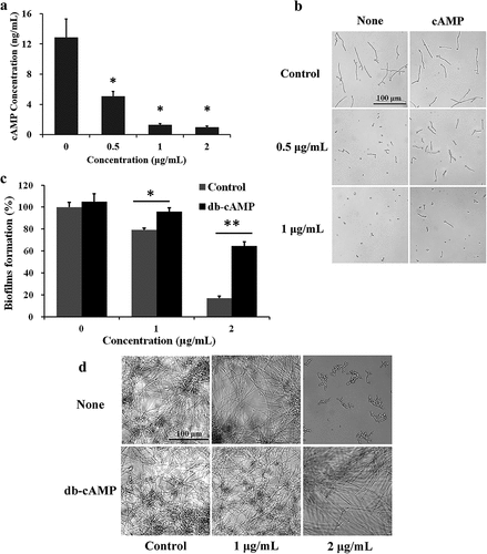
Figure 6. The effect of MPD on the farnesol secretion and related genes expression. (a) After farnesol was extracted from the supernatant of YEM30 strain treated with MPD, the content of farnesol was analysed by GC-MS. The fluorescence intensity of BWP17-DPP3-GFP strain was detected to reflect the expression of Dpp3. The intensity of fluorescence was monitored by fluorescence microscopy (b) and spectrofluorophotometry (c). (d) Qrt-PCR was used to detect the expression of hypha-related genes involved in Ras-Camp-Efg1 signalling pathway under MPD treatment. The bar in (b) indicates 50 µm. Results in (a), (c), and (d) are shown as means ± SDs.
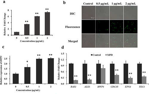
Figure 7. Effect of MPD on hyphal formation of DPP3 knockout mutant of C. albicans. SN152 and KWN2 strains were cultured in the RPMI 1640 medium containing different doses of MPD (0 and 1 μg/mL) at 37 °C. The imaging was observed by microscope every 3 h. The bar indicates 100 µm.
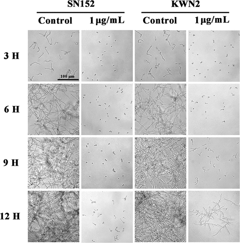
Table 3. The IC50 values of MPD against different cell lines.
Table 4. Hemolytic activity of MPD against human erythrocytes.
Figure 8. The effect of MPD on the G. mellonella-C. albicans infection model. (a) Healthy larvae were incubated with different doses of MPD for 5 days. (b) Larvae were infected with YEM30 strain and treated with PBS (control), FLC (2 μg/larva), or MPD (2 μg/larva) after 2 h of infection. Monitor larvae each day and calculate the survival rate. (c) The fungal burden of G. mellonella infected with C. albicans under different drug treatments was monitored every day after infection.
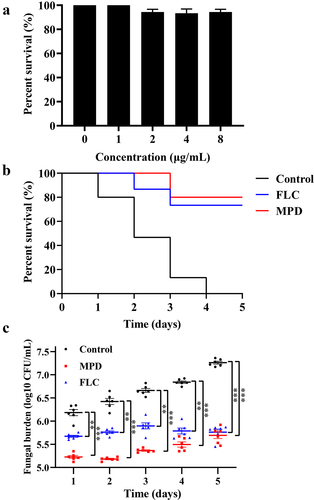
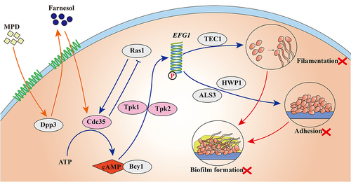
Figure 9. The underlying mechanism of MPD inhibiting virulence factors of C. albicans. MPD treatment increased the activity of Dpp3 in YEM30 strain to promote the secretion of farnesol. Farnesol directly inhibits the activity of Cdc35. Cdc35 converts ATP into cAMP, which combines with Bcy1, promoting transcription of downstream genes. These genes regulate a variety of virulence characteristics, such as the regulation of the hyphal morphology induced by EFG1 and TEC1. Meanwhile, EFG1 also regulates the adhesion induced by HWP1 and ALS3. Overall, MPD inhibited virulence factors through the Ras1-Camp-Efg1 pathway.
Data availability statement
The data that support the findings of this study are available at https://www.jianguoyun.com/p/DfLClH8Qn66aCxiR2ugEIAA.