
Figures & data
Figure 1. Mlkl-/- mice display increased susceptibility to S. pluranimalium pulmonary infection. Age- and sex-matched WT and Mlkl-/- mice (n = 10) were infected intranasally with 1 × 10⁹ CFUs S. pluranimalium strain STP1. (A, B) bacterial loads in the lung and BALF were determined at 24 h p.I. (C) total protein in BALF was assessed. (D) representative lung tissue structures were observed by H&E staining (upper panel, magnification × 100, lower panel, magnification, × 400). (E) representative TUNEL staining of apoptotic cells in the lung tissue (magnification, × 400). (F, G) representative immunohistochemical staining of F4/80 (a macrophages marker) and gr-1 (a neutrophil marker) were detected in lung sections (upper panel, magnification × 100, lower panel, magnification, × 400). (H) level of KC in lung homogenate supernatant was determined. (I) survival was monitored up to 72 h p.I. (n = 8 each group). Graphs are means ± standard deviation (SD) from data pooled 10 (A and B), five (C) or six (H) biological replicates. Statistical significance is indicated by *p < 0.05, **p < 0.01.
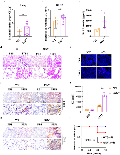
Figure 2. Mlkl-/- mice display increased susceptibility to S. pluranimalium systemic infection. Age- and sex-matched WT and Mlkl-/- mice (n = 10) were infected i.V. with 1 × 10⁸ CFUs S. pluranimalium strain STP1. (A-D) bacterial burden in blood, kidney, liver and spleen were quantitated at 24 hpi. (E) representative kidney tissue structures were observed by H&E staining (upper panel, magnification × 100, lower panel, magnification, × 400). (F) representative TUNEL staining of apoptotic cells in the kidney tissue (magnification, × 400). (G, H) representative immunohistochemical staining of F4/80 (a macrophages marker) and gr-1 (a neutrophil marker) were detected in kidney sections (upper panel, magnification × 100, lower panel, magnification, × 400). Graphs are means ± SD from data pooled 10 (A-D) biological replicates. Statistical significance is indicated by *p < 0.05, **p < 0.01.
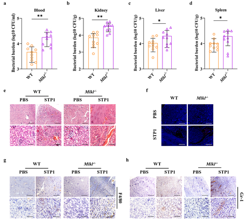
Figure 3. Inflammasome signalling is required for MLKL-mediated host protection against S. pluranimalium infection. Age- and sex-matched WT and Mlkl-/- mice were infected intranasally with log-phase S. pluranimalium strain STP1 (1 × 10⁹ CFUs) for 24 h. (A) lung tissues were collected, homogenized, and then immunoblotting for Caspase-1, IL-1β and GAPDH. Left, representative immunoblotting for Caspase-1 and IL-1β in lung tissues. Right, amounts of Caspase-1 and IL-1β determined by densitometry of protein bands from three experiments. The GAPDH served as a loading control. (B, C) levels of IL-1β and TNF-α in lung were determined. For one group of Mlkl-/- mice, 1.0 μg exogenous rIL-1β was injected intraperitoneally daily starting the day prior to the intranasally (1 × 10⁹ CFUs) or i.V. (1 × 10⁸ CFUs) inoculated with S. pluranimalium strain STP1 (n = 8 each group, at 24 h p.I.). (D-G) bacterial loads in the lung, BALF, blood and kidney were determined. (H, I) representative lung and kidney tissue structures were observed by H&E staining (upper panel, magnification × 100, lower panel, magnification, × 400). Graphs are means ± SD from data pooled six (B-C) or eight (D-G) biological replicates. Statistical significance is indicated by *p < 0.05, **p < 0.01.
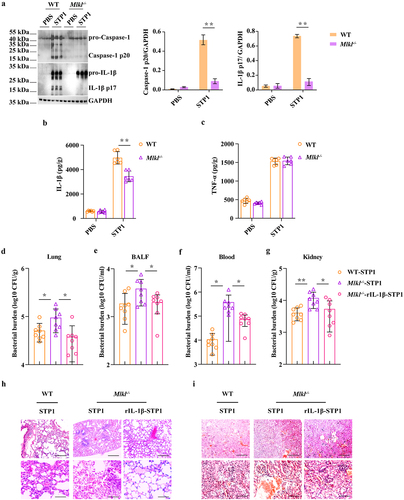
Figure 4. Mlkl deficiency suppresses NLRP3 inflammasome signalling. Age- and sex-matched WT and Mlkl-/- mice were infected intranasally (1 × 10⁹ CFUs) or i.V. (1 × 10⁸ CFUs) with S. pluranimalium strain STP1 for 24 h. (A, B) representative immunohistochemical staining of p-MLKL was performed in lung and kidney sections (upper panel, magnification × 100, lower panel, magnification, × 400). LPS-primed WT, Mlkl-/- and Nlrp3-/- BMDMs were incubated with or without potassium chloride for 30 min, followed by treatment with ATP (5 mM, 30 min) or S. pluranimalium strain STP1 (MOI = 50, 5 h). (C) top, representative images of immunoblotting for p-MLKL in cell lysates. Bottom, amounts of p-MLKL were quantified from three experiments. The GAPDH served as a loading control. (D, G and J) 0Left, representative immunoblotting for Caspase-1 and IL-1β in cell supernatants and cell extracts. The GAPDH served as a loading control. Right, amounts of Caspase-1 and IL-1β determined by densitometry of protein bands from three experiments. The cells with no treatment (NT) were taken as the control group. (E, F, H, I, K and L) culture supernatants were analysed for IL-1β and TNF-α by ELISA. Graphs are means ± SD from data pooled six (E, F, H, I, K and L) biological replicates. Statistical significance is indicated by *p < 0.05, **p < 0.01.
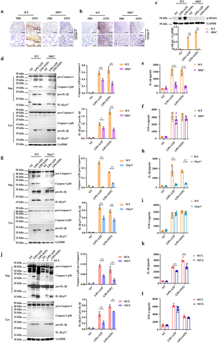
Figure 5. Nlrp3 deficiency impairs bacterial clearance and is detrimental to the host protection. Age- and sex-matched WT, Mlkl-/-and Nlrp3-/- mice (n = 10) were inoculated intranasally (1 × 10⁹ CFUs) or i.V. (1 × 10⁸ CFUs) with S. pluranimalium strain STP1 for 24 h. (A-F) bacterial burden in the lung, BALF, blood, kidney, liver and spleen were quantitated. (G, H) representative lung and kidney tissue structures were observed by H&E staining (upper panel, magnification × 100, lower panel, magnification, × 400). (I, J) representative TUNEL staining of apoptotic cells in the lung and kidney tissue (magnification, × 400). (K) lung tissues were collected, homogenized, and then immunoblotting for Caspase-1, IL-1β and GAPDH. Left, representative images of immunoblotting for Caspase-1 and IL-1β in lung tissues of WT and Nlrp3-/- mic. Right, amounts of Caspase-1 and IL-1β determined by densitometry of protein bands from three experiments. The GAPDH served as a loading control. (L, M) levels of IL-1β and TNF-α in lung were determined. LPS-primed WT, Mlkl-/- and Nlrp3-/- BMDMs were incubated with rIL-1β (1000 pg/ml) or PBS for 1 h before challenged with S. pluranimalium strain STP1 (MOI = 25, 6 h). (N, O) the supernatants were collected and plated on TSB agar plates to enumerate the bacteria. Graphs are means ± SD from data pooled 10 (A-F) or six (L-O) biological replicates. Statistical significance is indicated by *p < 0.05, **p < 0.01.
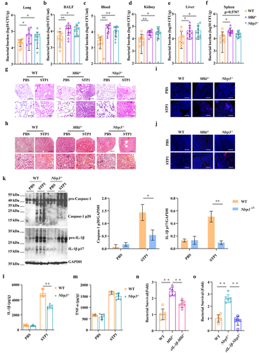
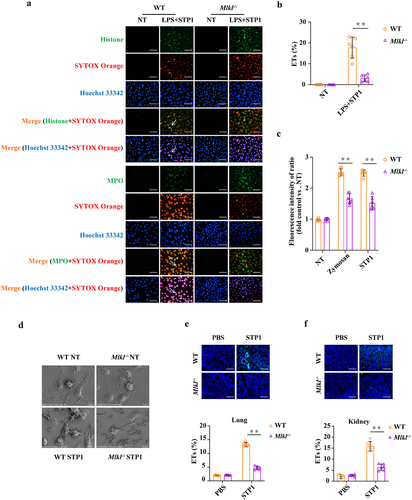
Figure 6. Mlkl deficiency causes a reduction of S. pluranimalium-triggered extracellular trap formation. LPS-primed WT and Mlkl-/- BMDMs were stimulated with S. pluranimalium strain STP1 (MOI = 25, 100 min). (A) representative immunofluorescence staining of METs formation as indicated by DNA decorated with histone or MPO within the ETs structures. Histone (green), MPO (green) and DNA (orange/blue). magnification, × 400. (B) METs were quantified by Fiji and presented as the percentage of ETs. (C) quantification of bacteria-induced METs formation using SYTOX green. Zymosan (1 mg/ml) stimulated cells were used as positive control. (D) the METs release was detected by SEM. Age- and sex-matched WT and Mlkl-/- mice were infected intranasally (1 × 10⁹ CFUs) or i.V. (1 × 10⁸ CFUs) with S. pluranimalium strain STP1 for 24 h. (E, F) representative immunofluorescence staining of histone was performed in lung and kidney sections, and ETs were quantified by Fiji and presented as the percentage of ETs. Graphs are means ± SD from data pooled six (B, C, E and F) biological replicates. Statistical significance is indicated by *p < 0.05, **p < 0.01.
Supplemental Material
Download MS Word (1.4 MB)Data Availability statement
The data supporting the findings of this study are available from the corresponding author upon reasonable request.