
Figures & data
Figure 1. Structure and genomic organization of SARS-CoV-2. The virion (a) consists of an unsegmented, positive ssRNA genome complexed with the nucleocapsid protein (n). Envelope-associated structural proteins include the trimeric spike (s), membrane (m), and envelope (e) proteins. Open reading frames (ORF) 1a and 1b are directly translated into the numerous nonstructural proteins (nsps) that form the replicase-transcriptase complex, including the RdRp. The remaining structural and accessory proteins (3a through 10) are derived from subgenomic mRnas (b). For additional information on coronavirus replication strategies, please see ref [Citation9].
![Figure 1. Structure and genomic organization of SARS-CoV-2. The virion (a) consists of an unsegmented, positive ssRNA genome complexed with the nucleocapsid protein (n). Envelope-associated structural proteins include the trimeric spike (s), membrane (m), and envelope (e) proteins. Open reading frames (ORF) 1a and 1b are directly translated into the numerous nonstructural proteins (nsps) that form the replicase-transcriptase complex, including the RdRp. The remaining structural and accessory proteins (3a through 10) are derived from subgenomic mRnas (b). For additional information on coronavirus replication strategies, please see ref [Citation9].](/cms/asset/b2d6243c-2d37-4cc4-a0ef-c2675963df75/kvir_a_2316438_f0001_oc.jpg)
Figure 2. Lung pathology in mice infected with SARS-CoV-2. a-c K18-hACE2 mouse infected with 5 × 10⁴ pfu SARS-CoV-2 WA10 that died on day 7. At low magnification (a), approximately 70% of the lung is hypercellular and there are multiple discrete foci of inflammation (arrows). The normal pulmonary architecture is obscured due to haemorrhage (arrow), oedema (stars), and inflammation (b). The alveolar and bronchiolar exudate is largely composed of neutrophils and there is rare type II pneumocyte hyperplasia (arrow) (c). (d-f) 6-week-old BALB/c mouse infected with 1 × 10⁴ pfu SARS-CoV-2 MA10 that was euthanized on day 4. There is minimal change at low magnification (d). There are occasional areas of hypercellularity (e) where septa are mildly thickened due to congestion and increased lymphocytes and macrophages (f).
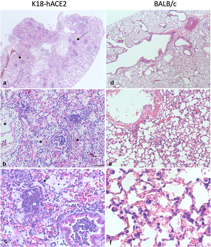
Table 1. Summary of animal models of COVID-19.
Data Availability statement
Data reported in the figure and table in this article have been deposited in a recognized data repository (Figshare.com) with a digital object identifier: 10.6084/m9.figshare.24348958