Figures & data
Figure 1. A schematic representation of the EV-A71 genome and current understanding of the picornaviruses 3Dpro protein. The enterovirus 5” end is linked to a VPg followed by an IRES. After translation of the single OFR, the polyprotein is cascade cleaved to form VP1, VP2, VP3, VP4, 2Apro, 2B, 2C, 3A, 3B, 3Cpro, and 3Dpol. at the 3” end of the EV-A71 genome is a polyA sequence. 3Dpol proteins catalyse viral RNA synthesis, catalyse VPg uridylation, interact with host proteins, and act as drug targets.
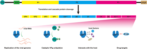
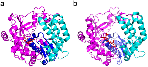
Figure 2. Structures of the RdRps of picornaviruses. a: EV-A71 3Dpol protein (PDB: 3N6L); b: EV-D68 3Dpol protein (PDB: 6L4R); c: CVA16 3Dpol protein (PDB: 5Y6Z); d: CVB3 3Dpol protein (PDB: 3DDK); e: PV 3Dpol protein (PDB: 4K4S); and f: FMDV 3Dpol protein (PDB: 6S2L). The palm, thumb and finger subdomains are shown in blue, cyan and magenta, respectively.
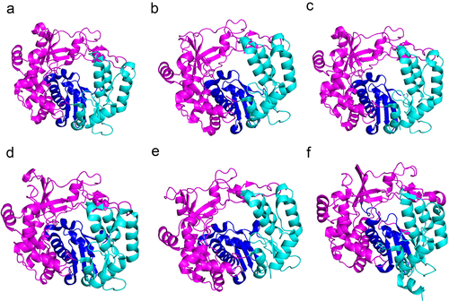
Figure 3. Diverse functions of the picornavirus 3Dpol protein and interactions with multiple host factors.
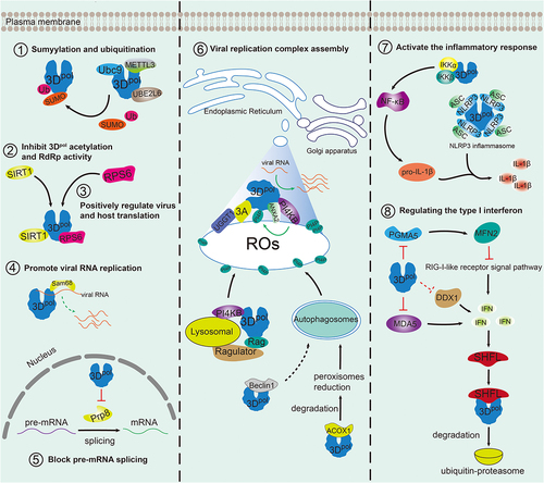
Figure 4. Structures of nucleoside/nucleotide analog inhibitors.
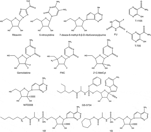
Table 1. RNA-dependent RNA polymerase nucleoside/nucleotide analog inhibitors.
Figure 5. Structures of non-nucleoside inhibitors.
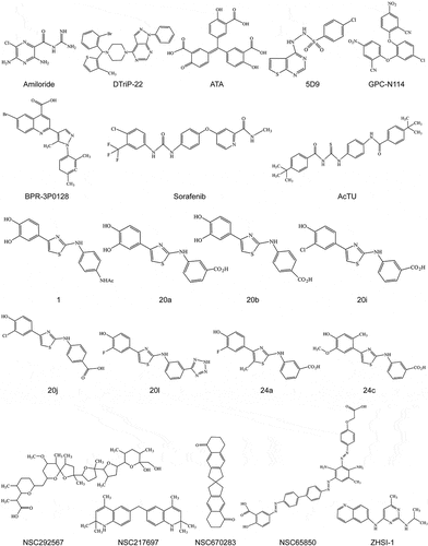
Table 2. RNA-dependent RNA polymerase non-nucleoside inhibitors.
Figure 6. X-ray crystal structure of 3Dpol and viral polymerase inhibitors. a: CVB3 3Dpol in complex with GPC-N114 (PDB: 4Y2A); b: EV-D68 3Dpol in complex with NADPH (PDB: 5ZIT). The palm, thumb and finger subdomains are shown in blue, cyan, and magenta cartoons, respectively. The inhibitors are shown in red sticks.