
Figures & data
Figure 1. Fig. 1 | STM infection caused death and intestinal lesions in the mice a: STM infection leads to decreased host weight in mice (n = 5). b: STM infection results in mortality of mice (n = 5). c: STM infection causes the shrinking of the small intestine and colon in mice. d: STM infection induces pathological changes in the small intestine and colon of mice. e: after STM infection, mice exhibit elevated expression of VILLIN, LGR5, and EpCAM proteins in the small intestine. f: following STM infection, there is a significant increase in the secretion of IL-22 protein and transcription of mIL-22 in the intestinal tract of mice.
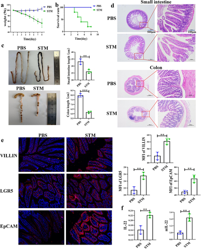
Figure 2. STM infection leads to significant changes in the gut microbiota and IL-22 secretion by ILC3 in mice.a: Changes in Lactobacillus Genus in the intestinal tract of piglets (n=5). b: Box plot of alpha diversity analysis (Chao1, Simpson index). c: Heatmap of beta diversity analysis. d: Inter-group PCA analysis. e: Differences in IL-22 secretion by CD45+ immune cells between the PBS and STM groups. f: Flow cytometry analysis of the number of ILC3 cells and the level of IL-22 secretion in the lamina propria of the mouse small intestine. g: Flow cytometry analysis of the number of TH17 cells and the level of IL-22 secretion in the lamina propria of the mouse small intestine.
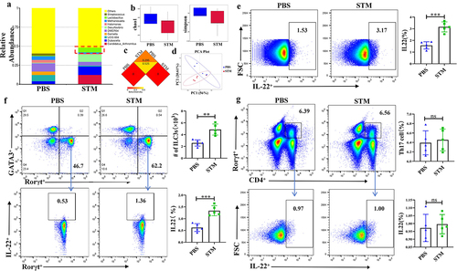
Figure 3. LGG can help mice resist STM infection.a: Feeding LGG significantly alleviates weight loss in mice after STM infection. b: Feeding LGG significantly reduces mortality in mice after STM infection. c: Feeding LGG significantly alleviates the shrinking of the small intestine and colon in mice after STM infection. d: Feeding LGG can significantly alleviate the pathological changes in the small intestine and colon in mice after STMinfection. e: Feeding LGG significantly reduces the expression of VILLIN, LGR5, and EpCAM proteins in the small intestine of mice after STM infection.
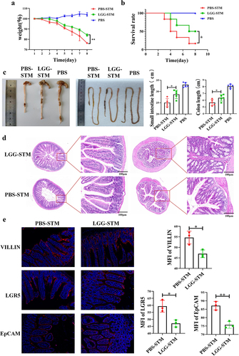
Figure 4. LGG promotes activation of ILC3 cells and is associated with IL-23 expression by TLR2 and DCs.a: Flow cytometry analysis of changes in ILC3 in the lamina propria of WT and KO mice after LGG feeding. b: Flow cytometry analysis of the level of IL-22 secretion by ILC3 in the lamina propria of WT and KO mice after LGG feeding. c: Flow cytometry analysis of the level of IL-23 expression by DCs in the lamina propria of WT and KO mice after LGG feeding.
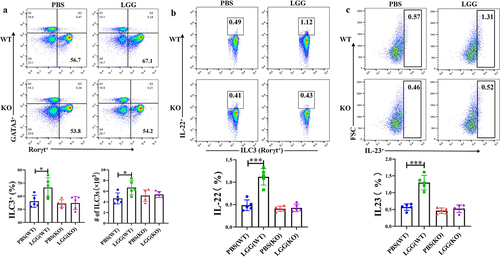
Figure 5. HK-LGG activates ILC3 by promoting IL-23 secretion from DCs.a: Flow cytometry analysis of the activating effect of HK-LGG, LGG supernatant, and DCs on IL-22 secretion by mouse ILC3. b: Transcription levels of the IL-22 gene in ILC3 under stimulation by different groups. c: Differential levels of IL-23 secretion promoted by HK-LGG and LGG supernatant from DCs.
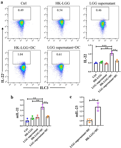
Figure 6. HK-LGG can promote the development of mice intestinal organoids through activating ILC3s to secrete IL-22.a: Crypts isolated from mice intestines were cultured in vitro, and the images depict the changes in organoid size over an 7-day period. b: Effects of HK-LGG; HK-LGG+DC; HK-LGG+ILC3 and HK-LGG+DC+ILC3 on the growth of intestinal organoids. Measure the size (surface area) and budding rate (building organoids) of the organs on the third and fifth days respectively. c: Immunofluorescence experiment demonstrating the expression of proteins such as Villin, EpCAM, LGR5 between different groups.
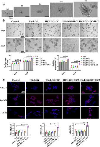
Figure 7. The pathway of HK-LGG enhances the intestinal mucosal immune barrier in mice.
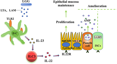
Fig._s1.jpg
Download JPEG Image (1.3 MB)
Fig._s3.jpg
Download JPEG Image (1.4 MB)
Fig._s2.jpg
Download JPEG Image (1.1 MB)
Data availability statement
The data that support the findings of this study are openly available in https://figshare.com/s/641045c425176601b3b6, DOI: 10.6084/m9.figshare.25826965. And 16S rRNA-seq data in https://www.ncbi.nlm.nih.gov/bioproject, reference number is [PRJNA1073044].