
Figures & data
Table 1. Primers used in the study.
Figure 1. Rescue of recombinant viruses. The HA titre of recombinant viruses.
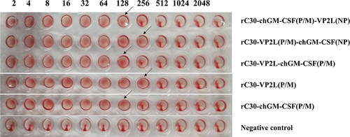
Figure 2. Identification of recombinant viruses. (a) RT-pcr-based detection of VP2L gene inserted into recombinant viruses: lane M: DL2000 marker; lane 1: rClone30-VP2L (P/M) virus; lane 2: rClone30-chGM-csf (P/M)-VP2L (NP) virus; lane 3: rClone30-VP2L (p/m)-chGM-csf (NP) virus; lane 4: rClone30-VP2L-chGM-csf (P/M) virus; lane 5: negative control. (b) RT-pcr-based detection of GM-CSF gene inserted into recombinant viruses: lane M: DL2000 marker; lane 1: rClone30-VP2L-chGM-csf (P/M) virus; lane 2: rClone30-chGM-csf (P/M)-VP2L (NP) virus; lane 3: rClone30-VP2L (p/m)-chGM-csf (NP) virus; lane 4: rClone30-chGM-csf (P/M) virus; lane 5: negative control. (c) Western blot-based detection of VP2L gene expression in recombinant viruses: lane 1: rClone30-VP2L-chGM-csf (P/M) virus; lane 2: rClone30-VP2L (p/m)-chGM-csf (NP) virus; lane 3: rClone30-VP2L (p/m)-chGM-csf (NP); lane 4: rClone30-VP2L (P/M) virus; lane 5: rClone30 virus; lane 6: culture medium control. (d) Western blot-based detection of GM-CSF gene expression in recombinant viruses: lane 1: rClone30-VP2L-chGM-csf (P/M) virus; lane 2: rClone30- chGM-csf (P/M)-VP2L (NP) virus; lane 3: rClone30-VP2L (p/m)-chGM-csf (NP) virus; lane 4: rClone30-chGM-csf (P/M) virus; lane 5: rClone30 virus; lane 6: culture medium control.
Figure 3. Growth characteristics of recombinant viruses. DF1 cells were evenly transfected with each virus at 0.1 MOI, and cell supernatants were collected at 24, 48, 72, and 96 h, and viral titre was calculated as TCID50 by the reed and Muench method.
Figure 4. Detection of anti-ndv and anti-ibdv antibody titre. (a) The anti-ndv antibody in the serum of chickens at 3, 5, 7, 10, 14, and 21 days after the first immunization was determined by HI test. (b) The anti-ibdv antibody in the serum of chickens at 7, 10, 14, and 21 days after the first immunization was measured by ELISA. Values are expressed as mean ±SD; **p < 0.01 versus rClone30-VP2L (P/M) virus-treated SPF chickens; #p < 0.05, ##p < 0.01 versus rClone30-chGM-csf (P/M) virus-treated SPF chickens; ^p < 0.05, ^^p < 0.01 versus commercial vaccine-treated SPF chickens.
Figure 5. Detection of inflammatory cytokines in the serum. The levels of IL-1β, IL-4, ifn-γ, and GM-CSF in the serum at 3, 5, 7, 10, 14, and 21 days after the first immunization were determined by ELISA. The chickens of the rClone30-VP2L (P/M) group, rClone30-chGM-csf (P/M) group, and commercial vaccine group were used as control. Values are expressed as mean ±SD; *p < 0.05, **p < 0.01 versus rClone30-VP2L (P/M) virus-treated SPF chickens; #p < 0.05, ##p < 0.01 versus rClone30-chGM-csf (P/M) virus treated SPF chickens; ^p < 0.05 versus commercial vaccine-treated SPF chickens.
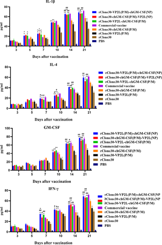
Figure 6. Immune response in immune cells. The percentages of CD4+ T, CD8+ T, B, and MHC-II+ cells were determined by FCM at 3, 5, 7, 10, 14 and 21 days after the first immunization. The chickens of the rClone30-VP2L (P/M) group, rClone30-chGM-csf (P/M) group and commercial vaccine group were used as controls. Values are expressed as mean ±SD; *p < 0.05, **p < 0.01 versus rClone30-VP2L (P/M) virus-treated SPF chickens; #p < 0.05, ##p < 0.01 versus rClone30-chGM-csf (P/M) virus-treated SPF chickens; ^p < 0.05, ^^p < 0.01 versus commercial vaccine treated SPF chickens.
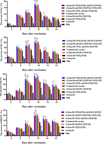
Figure 7. mRNA expression levels of inflammatory cytokines in white cells. The inflammatory cytokines IL-1β, ifn-γ, IL-4, and GM-CSF were analysed in the white cells by real-time PCR at 3, 5, 7, 10, 14, and 21 days post-first immunization. The chickens of the rClone30-VP2L (P/M) group, rClone30-chGM-csf (P/M) group, and commercial vaccine group were used as control. Values are expressed as mean ±SD; *p < 0.05, **p < 0.01 versus rClone30-VP2L (P/M) virus-treated SPF chickens; #p < 0.05, ##p < 0.01 versus rClone30-chGM-csf (P/M) virus-treated SPF chickens; ^p < 0.05 versus commercial vaccine-treated SPF chickens.
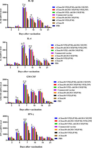
Figure 8. mRNA expression level of inflammatory cytokines in the spleen. The inflammatory cytokines IL-1β, IL-4, ifn-γ, and GM-CSF were analysed in the spleen by real-time PCR at 3, 5, 7, and 14 days post-first immunization. The chickens of the rClone30-VP2L (P/M) group, rClone30-chGM-csf (P/M) group, and commercial vaccine group were used as control. Values are expressed as mean ±SD; *p < 0.05, **p < 0.01 versus rClone30-VP2L (P/M) virus treated SPF chickens; #p < 0.05, ##p < 0.01 versus rClone30-chGM-csf (P/M) virus treated SPF chickens.
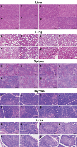
Figure 9. Histological structure of bursa, liver, lung, thymus, and spleen post immunization of SPF chickens. The bursa, liver, lung, thymus, and spleen samples were fixed by immersion in formalin, paraffin embedding, sectioned, and stained with haematoxylin and eosin. Histopathological changes were examined by light microscopy at a 200× field of view. Lane (a) PBS group; lane (b) rClone30 group; lane (c) rClone30-VP2L (P/M) group; lane (d) rClone30-chGM-csf (P/M) group; lane (e) rClone30-VP2L (p/m)-chGM-csf (NP) group; lane (f) rClone30-chGM-csf (P/M)-VP2L (NP) group; lane (g) rClone30-VP2L-chGM-csf (P/M) group; lane H: commercial vaccine group.
Data availability statement
All data generated or analyzed during this study are deposited in figshare, DOI: https://doi.org/10.6084/m9.figshare.26134012.