Figures & data
Figure 1. Normal cells induce RasV12 cell repulsion and increased contractility at the single cell level. Confocal images of coculture assays. Upper panels: Normal MDCK cells prelabeled with cell tracker dye (CMDRA) are added to confluent monolayers of GFP-RasV12 cells. Middle panels: GFP-RasV12 cells prelabeled with cell tracker dye (CMDRA) are added to confluent monolayers of GFP-RasV12 cells. Lower panels: GFP-RasV12 cells prelabeled with cell tracker dye (CMDRA) are added to confluent monolayers of normal MDCK cells. Cells were fixed 24 h after addition of prelabeled cells and stained with phalloidin (gray) and Hoechst (blue). Scale bar, 20 μm.
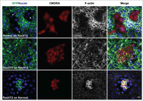
Figure 2. EphA2 receptor expressed on RasV12 cells is required to drive RasV12 cell repulsion and contractility following interaction with normal cells. Confocal images of coculture assays. Upper panels: Normal MDCK cells prelabeled with cell tracker dye (CMDRA) are added to confluent monolayers of GFP-RasV12 cells constitutively expressing EphA2 shRNA. Middle panels: GFP-RasV12 cells prelabeled with cell tracker dye (CMDRA) are added to confluent monolayers of GFP-RasV12 cells constitutively expressing EphA2 shRNA. Lower panels: GFP-RasV12 cells constitutively expressing EphA2 shRNA prelabeled with cell tracker dye (CMDRA) are added to confluent monolayers of normal MDCK cells. Cells were fixed 24 h after addition of prelabeled cells and stained with phalloidin (gray), anti-EphA2 antibody (magenta) and Hoechst (blue). XY images represent maximum projections of z-stacks. Scale bar, 20 μm.
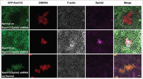
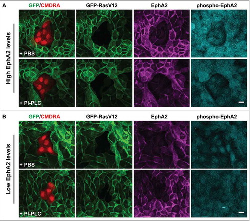
Figure 3. RasV12 cell repulsion and contractility is dependent on endogenous EphA2 expression levels and not on coexpressed ephrin-(A)ligands. Confocal images of coculture assays. (A) Normal MDCK cells prelabeled with cell tracker dye (CMDRA) are added to confluent monolayers of GFP-RasV12 cells that expressed high levels of EphA2 and were pretreated with either PBS (upper panels) or phosphatidylinositol-specific phospholipase C (PI-PLC) (lower panels). (B) Normal MDCK cells prelabeled with cell tracker dye (CMDRA) are added to confluent monolayers of GFP-RasV12 cells that expressed low levels of EphA2 and were pretreated with either PBS (upper panels) or phosphatidylinositol-specific phospholipase C (PI-PLC) (lower panels). Cells were fixed 24 h after addition of prelabelled cells and stained with anti-EphA2 (magenta), anti-phosphorylated EphA2 (Y594) (phospho-EphA2; cyan) and Hoechst (blue). XY images represent maximum projections of z-stacks. Scale bar, 20 μm.