Figures & data
Figure 1. Net1 deletion impairs RhoA activation in mouse bone marrow macrophages. (A) Net1 mRNA expression in M-CSF differentiated mouse bone marrow macrophages (BMMs). (B, C) Representative examples of F4/80 and CD68 expression in wild type (B) and Net1 KO (C) BMMs. (D) Quantification of the percent of cells with CD68 and F4/80 expression in differentiated BMMs. (E) Representative example of RhoA activity in BMMs, as measured by GST-RBD pulldown. (F) Quantification of RhoA activity in BMMs. For all plots, bars = median values. n.s. = not significant. ** = p < 0.01. *** = p < 0.001.
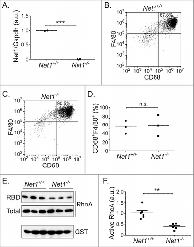
Figure 2. Net1 deletion reduces F-actin content in BMMs. (A) Representative images of pMLC2 staining of BMMs from wild type and Net1 KO mice. Scale bar is 10 µm. (B) Quantification of pMLC2 staining in multiple fields from three different BMM isolates for each genotype. (C) Western blot for pS19-MLC2 and total MLC2 in three different isolates of wild type and Net1 knockout BMMs. (D) Quantification of pS19-MLC2/MLC2 Western blots. (E) Representative images of F-actin staining in BMMs from wild type and Net1 KO mice. Scale bar is 10 µm. (F) Quantification of F-actin staining in multiple fields from three different BMM isolates for each genotype. (G) Western blot for pS3 cofilin and total cofilin in three different isolates of wild type and Net1 knockout BMMs. (H) Quantification of pS3-cofilin/cofilin Western blots. Bars are median values for all plots. n.s. = not significant. * = p < 0.05. *** = p < 0.001.
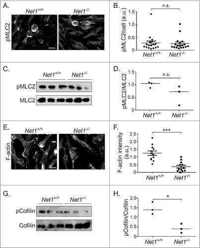
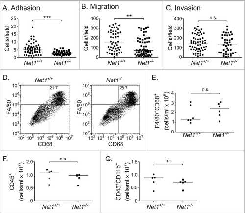
Figure 3. Net1 deletion impairs BMM adhesion and motility, but does not affect Matrigel invasion or peritoneal recruitment. (A) Adhesion of wild type and Net1 KO BMMs to fibronectin 30 minutes after plating. (B) Migration of wild type and Net1 KO BMMs in transwell inserts with MCP-1 in the bottom well. (C) Invasion of wild type and Net1 KO BMMs through Matrigel coated transwell inserts with MCP-1 in the bottom well. For all experiments, three different BMM isolates were assayed in triplicate for each genotype. (D) Representative examples of F4/80 and CD68 expressing cells in peritoneal isolates from wild type and Net1 KO mice three days after thioglycollate injection. (E, F, G) Quantification of F4/80+CD68+ (E), CD45+ (F), and CD45+CD11b+ cells in peritoneal isolates from thioglycollate injected wild type and Net1 KO mice. For all plots, bars = median values. n.s. = not significant; ** = p < 0.01; *** = p < 0.001.