Figures & data
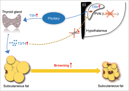
Figure 1. Schematic diagram of actions of LXRβ in controlling of thyroid hormone feedback in the brain and browning of SAT. TRH expressed by the neurons in PVN of the hypothalamus stimulates release of TSH from the anterior pituitary, which in turn stimulates thyroid hormone synthesis at the thyroid gland. T4/T3 in the circulation enter their target organs such as SAT and activate the browning process. In reverse, the circulating thyroid hormones negatively regulate their own production through targeting both pituitary and hypothalamus. LXRβ is able to transcriptionally inhibit the expression of TRH in the PVN area. Genetic depletion of LXRs releases their transcriptional suppression on TRH and breaks the negative feedback loop due to the lack of TRs in the PVN area, thus promotes TSH secretion in the pituitary and activates the synthesis of thyroid hormone, which eventually increases the browning of SAT.