
Figures & data
Figure 1. Mature adipocytes rapidly internalize a large amount of isolated mitochondria. (a) Mature immortalized murine brown adipocytes were incubated with DsRed2-labelled mitochondria derived from the same cell line for 24 hours. Experiment was reproduced twice. Scale bars 100 µm. (b) Time course of mitochondrial transformation into mature immortalized murine brown adipocytes. Recipient cells were incubated in the presence of murine brown adipocyte-derived DsRed2-labelled mitochondria (1 mg/ml) for indicated amounts of time. (c) Capacity of mature immortalized murine brown adipocytes to integrate mitochondria. Recipient cells were incubated in the presence of indicated amounts of DsRed2-labelled mitochondria derived from murine brown adipocytes for 24 hours, washed 5 times with PBS and fluorescence was measured after another 24 hours. Data is normalized to the protein content and expressed as mean ± SEM. N = 6 from 2 independently performed experiments.
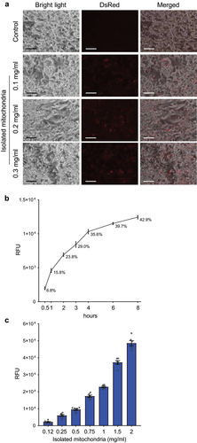
Figure 2. Internalized mitochondria remain functional in recipient mature immortalized brown and human white adipocytes for several days. (a) Representative western blot and (b) quantification of UCP1 protein, (c) mitochondrial respiration and (d) extracellular acidification rate in recipient mature immortalized murine brown adipocytes 24 hours after mitochondrial transformation. (e) Representative western blot and (f) quantification of UCP1 protein, (g) mitochondrial respiration and (h) extracellular acidification rate in recipient hMADS cells differentiated into mature white adipocytes 96 hours after transformation of human brown adipocyte-derived mitochondria. Data is expressed as mean ± SEM. N = 6 from 2 independently performed experiments. Statistical significance was calculated using ANOVA and is indicated as: *p < 0.05; **p < 0.01; ***p < 0.001.
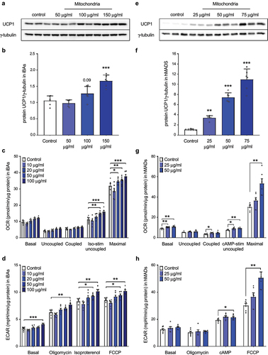
Figure 3. Internalized mitochondria do not trigger transcriptional changes in recipient mature adipocytes. (a) Expression of nuclear-encoded brown/beige adipocyte markers and (b) mitochondrially encoded genes in recipient white hMADS cells transformed with mitochondria isolated from human brown adipocytes (n = 4). (c) Glycerol release into media by immortalized murine brown adipocytes transformed with exogenous mitochondria derived from the same cell line (n = 6). (d) Oxygen consumption rate and (e) extracellular acidification rate in human white adipocytes 8 days after transformation of human brown adipocyte-derived mitochondria. Data is expressed as mean ± SEM. Experiments were reproduced twice. Statistical significance was calculated using ANOVA and T-test.
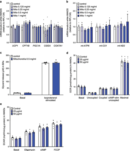
Figure 4. Administration of exogenous mitochondria into inguinal WAT does not increase energy expenditure in mice in vivo. Administration of immortalized murine brown adipocyte-derived mitochondria does not affect (a) whole-body energy expenditure, (b) oxygen consumption, (c) respiratory exchange ratio (RER), (d) food and (e) water intake or (f) locomotor activity in C57BL/6 mice (n = 5). Data is expressed as mean ± SEM. Statistical significance was calculated using a T-test.
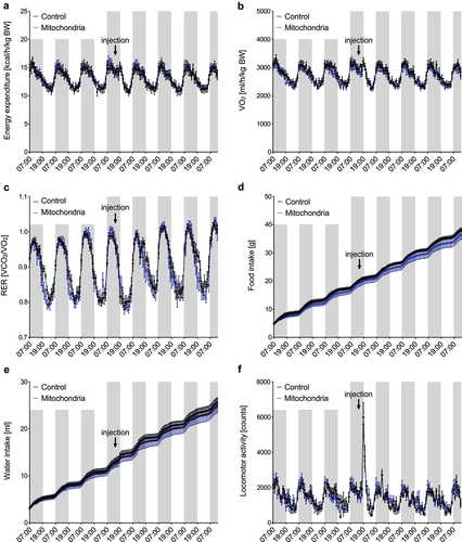
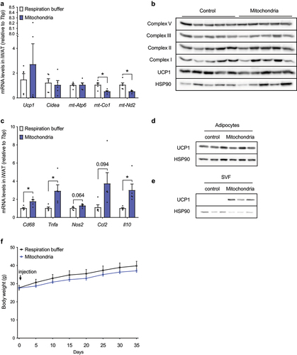
Figure 5. Administration of exogenous mitochondria neither affects UCP1 and mitochondrial content in inguinal WAT, nor body weight gain under obesogenic conditions in vivo. (a) Expression of thermogenic and mitochondrially encoded genes in iWAT of C57BL/6 mice 6 days after injection of respiration buffer or freshly isolated brown adipocyte-derived mitochondria (20 mg; 10 mg per depot; n = 5). (b) Immunostaining of UCP1 and individual OXPHOS complexes in iWAT of C57BL/6 mice 6 days after injection of respiration buffer or isolated brown adipocyte-derived mitochondria (20 mg; 10 mg per depot; n = 5). (c) Expression of inflammatory gene markers in iWAT of mice 6 days after injection of respiration buffer or freshly isolated brown adipocyte-derived mitochondria (20 mg; 10 mg per depot; n = 5). UCP1 protein content in adipocyte-enriched fraction and (e) stromal vascular fraction of inguinal white adipose tissue 48 hours after administration of immortalized murine brown adipocyte-derived mitochondria (1 mg per depot). (f) Body weight gain in mice injected with respiration buffer or isolated brown adipocyte-derived mitochondria (15 mg per iWAT depot) on high fat diet (n = 5). Data is expressed as mean ± SEM. Statistical significance was calculated using a T-test and is indicated as: *p < 0.05.
Data availability statement
All data presented in the manuscript are available under: https://data.mendeley.com/datasets/pwgc8z9zdm/2