Figures & data
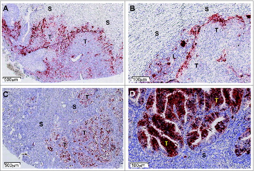
Figure 1. IDO1 Protein expression in human tumors assessed by immunohistochemistry. Illustrative images from formalin-fixed paraffin-embedded tissue microarray sections of cervical (A, B) and endometrial carcinomas (C, D) stained with the anti-IDO1 antibody 4.16H1. Tumoral (T), stromal (S), and lymphocyte-enriched (L) areas are indicated. Immunolabeled cells are stained dark red.