Figures & data
Figure 1 (see previous page). Akt inhibition preserves the TCM phenotype and enhances the proliferative ability of CD8+ T cells. Non-fractionated splenocytes from pMel-1 mice were stained with VCT and activated with gp10025–33 peptide (1 µmol/L) in the presence or absence of MK-2206 (0.67, 2, and 6 µmol/L). The concentration of the inhibitors was maintained throughout the experiment. The cells were re-stimulated with gp10025–33 peptide on days 7, 14, and 21 and their phenotype and proliferation were assessed. The gated cells were viable (7AAD-) CD8+Vβ13+. The data are representative of at least 4 independent experiments. (A) In this representative example, CD8+ T cells from a naïve spleen (far left) are mainly (72%) naïve cells (CD62LhiCD44lo). Sixty-seven percent (67%) of non–MK-2206-treated CD8+ T cells (third graph from left) are TEM cells (CD62LloCD44hi) and less than 1% are naïve cells (CD62LhiCD44lo). This changes when cells are treated with MK-2206 (far right), as 65% of the cells possess the TCM phenotype (CD62LhiCD44hi) and 14% are naïve cells (CD62LhiCD44lo). (B) After 3 d of stimulation, the proliferation of CD8+ T cells was inhibited in a dose-dependent manner by MK-2206 (VCT dilution) (far left). CD8+ T cells treated with MK-2206 expand at a significantly high rate with further stimulations (middle graph; data normalized to the non-treated control [GP100]). MK–2206-treated CD8+ T cells secrete significantly higher levels of IL-2 following stimulations 2 and 3, which is consistent with their higher proliferative potential (far right). *, P < 0.05; **, P < 0.01; ****, P < 0.0001. (C) Akt inhibition by MK-2206 maintains a high level of CD62L expression in CD8+ T cells on Day 3, and on Day 7 after each stimulation with gp100. (D) Akt inhibition by MK-2206 maintains high levels of CD127 in CD8+ T cells on Day 3, and on Day 7 after each stimulation with gp100. (E) Akt inhibition by MK-2206 inhibits the upregulation of KLRG-1 in CD8+ T cells after the second and third stimulations with gp100.
![Figure 1 (see previous page). Akt inhibition preserves the TCM phenotype and enhances the proliferative ability of CD8+ T cells. Non-fractionated splenocytes from pMel-1 mice were stained with VCT and activated with gp10025–33 peptide (1 µmol/L) in the presence or absence of MK-2206 (0.67, 2, and 6 µmol/L). The concentration of the inhibitors was maintained throughout the experiment. The cells were re-stimulated with gp10025–33 peptide on days 7, 14, and 21 and their phenotype and proliferation were assessed. The gated cells were viable (7AAD-) CD8+Vβ13+. The data are representative of at least 4 independent experiments. (A) In this representative example, CD8+ T cells from a naïve spleen (far left) are mainly (72%) naïve cells (CD62LhiCD44lo). Sixty-seven percent (67%) of non–MK-2206-treated CD8+ T cells (third graph from left) are TEM cells (CD62LloCD44hi) and less than 1% are naïve cells (CD62LhiCD44lo). This changes when cells are treated with MK-2206 (far right), as 65% of the cells possess the TCM phenotype (CD62LhiCD44hi) and 14% are naïve cells (CD62LhiCD44lo). (B) After 3 d of stimulation, the proliferation of CD8+ T cells was inhibited in a dose-dependent manner by MK-2206 (VCT dilution) (far left). CD8+ T cells treated with MK-2206 expand at a significantly high rate with further stimulations (middle graph; data normalized to the non-treated control [GP100]). MK–2206-treated CD8+ T cells secrete significantly higher levels of IL-2 following stimulations 2 and 3, which is consistent with their higher proliferative potential (far right). *, P < 0.05; **, P < 0.01; ****, P < 0.0001. (C) Akt inhibition by MK-2206 maintains a high level of CD62L expression in CD8+ T cells on Day 3, and on Day 7 after each stimulation with gp100. (D) Akt inhibition by MK-2206 maintains high levels of CD127 in CD8+ T cells on Day 3, and on Day 7 after each stimulation with gp100. (E) Akt inhibition by MK-2206 inhibits the upregulation of KLRG-1 in CD8+ T cells after the second and third stimulations with gp100.](/cms/asset/565bf607-4b96-43c7-aa07-f9e3adce5655/koni_a_1005448_f0001_c.jpg)
Figure 2. Akt inhibition by MK-2206 maintains a high level of IFNγ and TNF secretion in CD8+ T cells. CD8+ T cells from pMel-1 mice were stimulated with gp10025–33 peptide (1 µmol/L) in the presence or absence of MK-2206 (0.67 µmol/L). On days 7 and 14, CD8+ T cells were re-stimulated with gp10025–33 peptide and the IFNγ and TNF levels in the supernatant were assessed after 24 h using CBA. Granzyme B expression was assessed on days 7 and 14. The data are representative of at least 2 independent experiments. (A) The ability of CD8+ T cells to produce IFNγ with subsequent stimulations is significantly diminished. CD8+ T cells treated with MK-2206 maintain their ability to secrete IFNγ with further stimulations. *, P < 0.05; ****, P < 0.0001. (B) CD8+ T cells treated with MK-2206 produce significantly higher levels of TNF and maintain this ability with further stimulations. *, P < 0.05. (C) Following the first stimulations, all the cells produce Granzyme B. A higher percentage of CD8+ T cells treated with MK-2206 or Akt-1/2 inhibitor produce a high level of Granzyme B following the second stimulation.
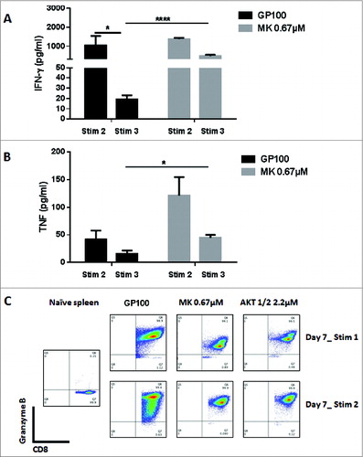
Figure 3 (see previous page). The Inhibition of Akt1 and Akt2 preserves TCM cells and enhances the proliferative ability of CD8+ T cells. Non-fractionated splenocytes from pMel-1 mice were stained with VCT and activated with gp10025–33 peptide (1 µmol/L) in the presence or absence of Akt-1/2 inhibitor (2.2, 6.7, and 20.1 µmol/L). The cells were re-stimulated with gp10025–33 on days 7, 14, and 21. The gated cells were viable (7AAD-) CD8+Vβ13+. (A) Akt1 and Akt2 inhibition preserves the TCM phenotype. In this representative example, 76% of non-treated CD8+ T cells are TEM cells (CD62LloCD44hi) and less than 1% are naïve cells (CD62LhiCD44lo), whereas CD8+ T cells treated with an Akt-1/2 inhibitor consist of 76% TCM cells (CD62LhiCD44hi) and 22% naïve cells (CD62LhiCD44lo). (B) The proliferation of CD8+ T cells is inhibited by the Akt-1/2 inhibitor in a dose-dependent manner (Day 3). The expansion of CD8+ T cells treated with the inhibitor is significantly enhanced with further stimulations. Data are normalized to the non-treated control (GP100). *, P < 0.05. (C) Akt1 and Akt2 inhibition maintains a high level of CD62L on Day 3, and on Day 7 after each stimulation. (D) Akt1 and Akt2 inhibition maintains a high level of CD127 on Day 3, and on Day 7 after each stimulation. (E) Akt1 and Akt2 inhibition mitigates the upregulation of KLRG-1 in CD8+ T cells after the second and third stimulations. Following the third stimulation, the KLRG-1 level was dramatically increased and, therefore, the biexponential scale of the graph had to be adjusted for presentation purposes.
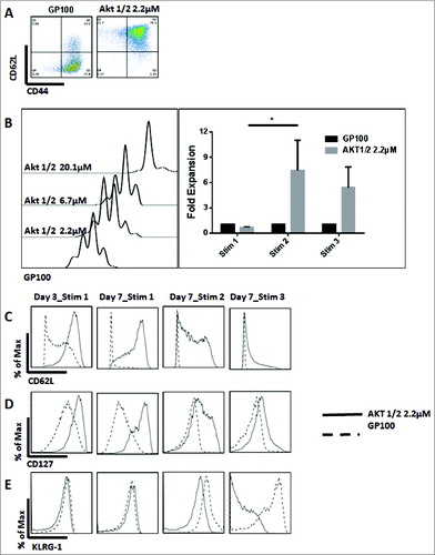
Figure 4. The absence of Akt1 and Akt2 isoforms, but not Akt3, preserves the TCM phenotype. Enriched CD8+ T cells from Akt1, -2, and -3 KO and WT mice were stimulated with anti-CD3 (1 μg/mL) and co-stimulated with anti-CD28 (2.5 μg/mL) antibodies. The phenotype of the cells was assessed on Day 7. The gated cells were viable (7AAD-) CD8+. (A) In this representative example, WT CD8+ T cells consisted of 83% TEM cells (CD62LloCD44hi). Akt1 KO CD8+ T cells consisted of 55% TCM cells (CD62LhiCD44hi), with 43% of the cells being TEM cells. Akt2 KO CD8 T cells consist of 36% TCM and 63% TEM cells. Akt3 KO CD8+ T cells consisted of 86% TEM cells, whereas only 12% were TCM cells. (B) Akt1 KO cells express a higher level of CD62L than Akt2 KO cells, which in turn express higher levels of these markers than WT and Akt3 KO cells that express similar levels. (C) Akt1 KO cells express a higher level of CD127 than Akt2 KO cells, which in turn express higher levels of these markers than WT and Akt3 KO cells that express similar levels.
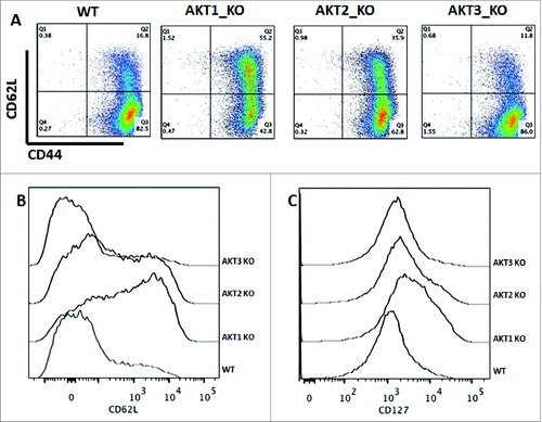
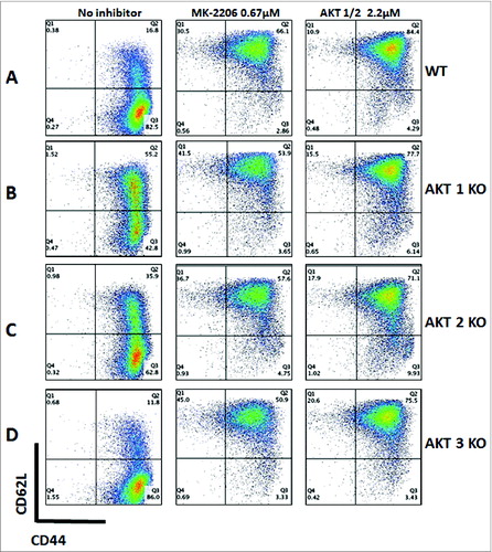
Figure 5. Akt inhibition preserves the TCM phenotype in WT and Akt KO mice. Enriched CD8+ T cells from Akt1, -2, and -3 KO and WT mice were stimulated anti-CD3 (1 μg/mL) and co-stimulated with anti-CD28 (2.5 μg/mL) antibodies in the presence or absence of MK-2206 (0.67 µmol/L) or an Akt-1/2 inhibitor (2.2 μmol/L). The phenotype of the cells was assessed on Day 7. The gated cells were viable (7AAD-) CD8+. (A) Untreated CD8+ T cells from WT mice consist mainly of TEM cells, whereas those treated with MK-2206 or Akt-1/2 inhibitors consist mainly of TCM cells. (B) CD8+ T cells from Akt1 KO mice possess significantly more TCM cells than WT without any inhibitors. Once treated with MK-2206 or Akt-1/2 inhibitors, more TCMcells (with a higher CD62L expression) are maintained comparable to treated WT cells. (C) CD8+ T cells from Akt2 KO mice possess significantly more TCM cells than WT without any treatments, although less than that observed from Akt1 KO mice. Treatment with MK-2206 or Akt-1/2 inhibitors maintains a significantly higher percentage of TCM cells comparable to WT and Akt1 KO treated cells. (D) Similar to WT, CD8+ T cells from Akt3 KO mice consist mainly of TEM cells. Treatment with MK-2206 or Akt-1/2 inhibitors maintains a significantly higher percentage of TCM cells comparable to WT and Akt1 and -2 KO treated cells.