
Figures & data
Figure 1. Antitumor activity of 5-AZA-CdR combined with mAb 9H10 in the syngeneic TS/A mouse tumor model. BALB/c mice were inoculated sc with 2 × 10⁵ TS/A cells. Groups of five mice were injected ip with 5-AZA-CdR on days 0, 7 (1st cycle) and 42, 49 (2nd cycle); with mAb 9H10 on days 2, 5, 8 (1st cycle) and 44, 47, 50 (2nd cycle); with combined administration of 5-AZA-CdR and mAb 9H10 according to the above reported schedules, or with saline solution for control. Tumor volumes from mice were measured periodically, all along the treatment. Mean tumor volume for each group are reported. Vertical arrows indicate days of different treatments. *, p ≤ 0.05; **, p ≤ 0.01; ***, p ≤ 0.001 vs. tumor volume of control group. †, p ≤ 0.05; ††, p ≤ 0.01 vs. tumor volume of 5-AZA-CdR group. Representative data are shown from three experiments.
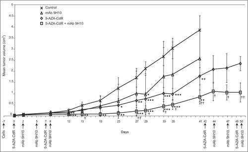
Figure 2. Regulation of Cancer Testis Antigen expression by 5-AZA-CdR combined with mAb 9H10 in the syngeneic TS/A mouse tumor model. (A) Total RNA was extracted from tumors excised from TS/A grafted mice treated with: saline solution, as control group (CTRL), mAb 9H10, 5-AZA-CdR, or the combination of 5-AZA-CdR with mAb 9H10. RT-PCR analysis was performed using P1A-, Mage-a- or β-actin-specific primer pairs. PCR products were then separated on a 2% agarose gel and visualized by ethidium bromide staining. Total RNA from mouse testis and splenocytes was utilized as positive (ctrl +) or negative (ctrl −) controls, respectively. Figure shows data from three representative mice out of five for control and treated groups. (B) genomic DNA was extracted from TS/A tumors excised from control mice (black) and mice treated with mAb 9H10 (white), 5-AZA-CdR (vertical line) or the combination of 5-AZA-CdR with mAb 9H10 (horizontal line). Real-time quantitative Methylation-Specific PCR analyses of P1A promoter were performed on bisulfite-modified genomic DNA, extracted from three out of five mice per group, using methylated- or unmethylated-specific primer pairs. Data are reported as percentage of average methylation that was defined as the ratio between methylated molecules and the sum of methylated and unmethylated molecules. Bars, SD; *, p ≤ 0.05 vs. control group.
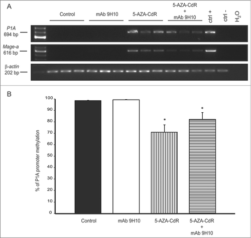
Figure 3. Modulation of MHC class I antigen expression and T cell infiltration by 5-AZA-CdR combined with mAb 9H10 in the syngeneic TS/A mouse tumor model. BALB/c mice were inoculated sc with 2 × 10⁵ TS/A cells. Groups of 5 mice were injected ip with 5-AZA-CdR; with mAb 9H10; with combined administration of 5-AZA-CdR and mAb 9H10 according to the above reported schedules, or with saline solution for control. A week after the end of treatment, neoplastic and normal tissues were excised and snap frozen in liquid nitrogen. Four micron acetone-fixed cryostat sections were processed for IHC assays. Representative results from tumors (A–F) and normal tissues (G–N) are reported. (A– C): MHC class I staining of tumors from mice treated with saline solution, with mAb 9H10 or 5-AZA-CdR, respectively; (D– F): CD3 staining of tumors from mice treated with saline solution, with mAb 9H10 or the combination of 5-AZA-CdR and with mAb 9H10, respectively; (G–I and L–N): CD3 staining of glandular epithelium of large intestine, liver, lung, myocardium renal parenchyma and dermis from mice treated with mAb 9H10 or 5-AZA-CdR, respectively. (A–F), 200× magnification, (G–M), 160× magnification; (N), 250× magnification. The arrowed marks the dermal-epidermal junction; black arrows, CD3+ lymphocytes.
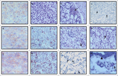
Figure 4. Antitumor activity of 5-AZA-CdR combined with mAb 9H10 in immunocompromised mice. Athymic nude mice (A) and SCID/Beige (B) mice were inoculated sc with 2 × 10⁵ TS/A cells. Groups of 4 mice, for each strains, were injected ip with 5-AZA-CdR on days 0 and 7; with mAb 9H10 on days 2, 5, 8; with combined administration of 5-AZA-CdR and mAb 9H10 according to the above reported schedules, or with saline solution for control. Tumor volumes from mice were measured periodically, all along the treatment. Mean tumor volumes for each group are reported. Vertical arrows indicate days of different treatments. *, p ≤ 0.05; **, p ≤ 0.01 vs. tumor volume of control group.
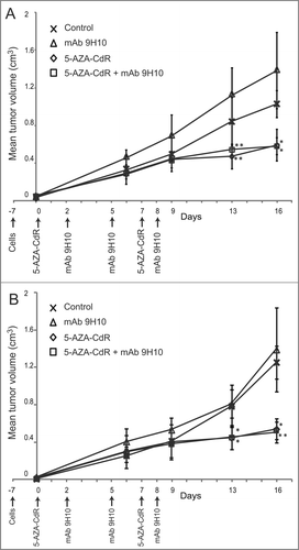
Table 1. Count of T cell infiltrates in neoplastic tissues from control and treated mice grafted with TS/A cells