Figures & data
Figure 1. Meriva reduced IL-6 production in 4T1 breast tumors. BALB/c mice were injected with 105 4T1 tumor cells in the mammary fat pad, and treated with various doses of Meriva (0, 5, 10, and 25 mg/mouse) orally (every 3 d for 14 d starting when tumors are 1–1.5 cm in diameter), and 2 d after the last treatment mice were euthanized and their tumors were homogenized and analyzed for the presence of IL-6 by ELISA (A). In addition the effect of Meriva was measured on tumor weight (B) and lung metastases (C). Representative of two experiments, n = 3 mice per group. The error bars represent the SEM. All groups were compared to the Saline group. Mann-Whitney test. *P < 0.05. Values P < 0.05 were considered statistically significant.
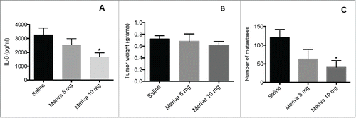
Figure 2. Cryoablation but not Meriva reduced the MDSC population in blood of 4T1 mice. BALB/c mice were injected with 105 4T1 tumor cells in the mammary fat pad and treated with cryoablation (20%) 14 d later. Meriva was administered (10 mg/300 μL Saline; orally), every 3 d for 14 d, starting on day 3 after cryoablation and then euthanized for analysis 2 d after the last treatment (day 30 after tumor cell injection). Myeloid-derived suppressor cells (MDSC)(CD11b+Gr1+) were analyzed in blood by flow cytometry and shown in flow cytometry profile (A) or averaged (B). The percentage of MDSC was determined in the total leucocyte population in blood. Representative of two experiments with n = 5 mice per group. The error bars represent the SEM. All groups were compared to cryoablation plus Meriva group. Mann-Whitney test. *P < 0.05. Values P < 0.05 were considered statistically significant.
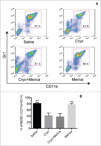
Figure 3. TAA-specific T cell responses generated through cryoablation were significantly improved by Meriva in 4T1 model. BALB/c mice were challenged with 4T1 tumor cells and treated with cryoablation plus Meriva as described in . Two days after the last treatment all mice were euthanized and analyzed for CD8+ T cell responses to TAA in vitro in the spleen by ELISPOT. CD8+ T cell responses to Mage-b (transfection of spleens of treated and control mice with pcDNA3.1-Mage plus pCMV-GM-CSF) (A), or to Survivin (transfection of spleens of treated and control mice with pcDNA3.1-Survivin plus pCMV-GM-CSF) (B) were analyzed, and a similar experiment was performed with or without anti-MHC Class I antibodies added to the wells as indicated in the figure and restimulated with pc-DNA3.1-Mage-b (C) or Survivin peptides (D). No restimulation was used as negative control (E). CD8+ T cells were depleted using magnetic beads technique. The results of two experiments were averaged with n = 5 mice per group. The error bars represent the SEM. All groups were compared to the cryoablation plus Meriva group. Mann-Whitney test. *P < 0.05, ** P < 0.01, ****P < 0.0001. Values P < 0.05 were considered statistically significant.
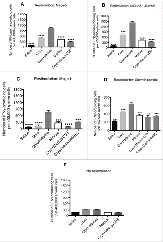
Figure 4. Cryoablation and Meriva significantly reduced metastatic breast cancer in 4T1 model. BALB/c mice were injected with 105 4T1 tumor cells in the mammary fat pad and treated with cryoablation (20% protocol) and Meriva as described in . All mice were euthanized 2 d after the last treatment and analyzed for tumor weight (A) and the number of metastases in the lung was counted using a preparation microscope (B). A representative example of a primary tumor and metastases in the lungs (perfused with Indian Ink) is depicted in C. The error bars represent the SEM. All groups were compared to the cryoablation plus Meriva group. Mann-Whitney test. *P < 0.05, *** P < 0.001. Values P < 0.05 were considered statistically significant.
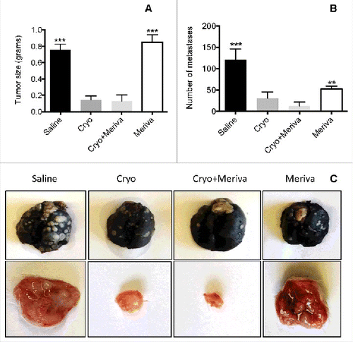
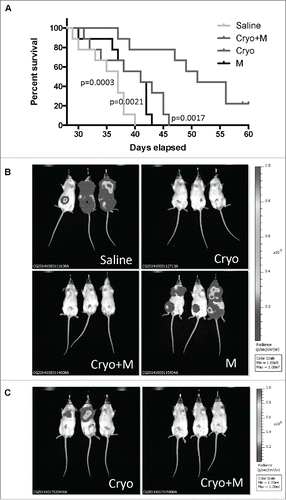
Figure 5. Cryoablation followed by Meriva significantly improved the survival rate and reduced the number of lung metastases. BALB/c mice were injected with 105 4T1 tumor cells in the mammary fat pad and treated with cryoablation (20% protocol) and Meriva as described in , but now maintained until they succumbed spontaneously (A). The only difference with is that in this survival and IVIS study Meriva was administered until the mice succumbed, instead of for 14 d only. Four groups were included – I: Saline; II: cryoablation; III: cryoablation and Meriva; and IV: Meriva. In this experiment n = 9 mice per group. Results were analyzed by Log-rank Mantel-Cox test. P < 0.05 is significant. All groups were compared to the cryoablation plus Meriva group. M = Meriva; Cryo = cryoablation. Median survival times: saline 37 d, cryoablation 41 d, cryoablation plus Meriva 51 d, and Meriva 41 d. In parallel, mice were injected with 105 4T1.2luc3 cells (same groups as in A), to analyze the effect of treatment live by the IVIS system (B). 4T1.2luc3 tumor cells grew more slowly then 4T1 cells. Therefore, Cryoablation was given on day 21 after tumor cell injection instead of day 14, and Meriva was administered 3 d after cryoablation. On day 38 all mice were monitored by IVIS. n = 3 mice per group. On day 58, when all mice in the saline group and Meriva group were dead the mice in the cryoablation group and in the cyroablation and Meriva group were still alive and monitored by in vivo imaging system (IVIS) (C). The total luciferase signal (tumor plus metastases) for each mouse was quantified on days 38 and 58 as shown in Supplemental Table 1.