Figures & data
Table 1. Patients' characteristics
Table 2. Cumulative survival according to patients' HLA-A genotype and HLA-G and E tumor cells expresssion
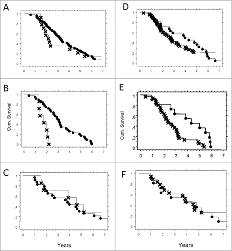
Figure 1. (A–F) Probability of survival correlated to expression of HLA-G and HLA-E, presented by Kaplan–Meier. Pathologic (X) or normal expression (•) in serous adenocarcinoma tumor cells. (A) HLA-G in the total cohort N.S. (B) HLA-G in worst prognosis group (HLA-A*02) p = 0.0003. (C) HLA-G in otherwise group (HLA-OW) N.S. (D) HLA-E in the total cohort p = 0.003. (E) HLA-E in worst prognosis group (HLA-A*02) p = 0.0003. (F) HLA-E in better prognosis group (HLA-OW) N.S.
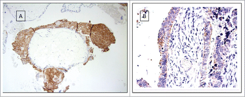
Figure 2. Tumor cells expressing HLA-G shows aberrant HLA-E expression and loss of classical MHC expression (A) HLA-G. (B) HLA-E, score 3. (C) Classical MHC class I HC.
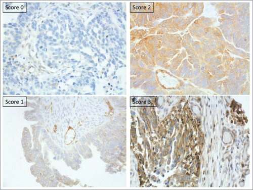
Figure 3. Cumulative survival by Kaplan–Meier analysis. All cases are HLA-G positive tumors with concordant loss of classical MHC. The worst prognosis was only significantly determined by HLA-A*02 genotype. HLA-A*02 (X), HLA-A otherwise (•). p = 0.003.
Table 3. Characteristic of malignant cells obtained from ascites
Table 4. Immunocomptentent infiltrating cells
Figure 4. Cumulative survival by Kaplan–Meier analysis. HLA-A*02 patients with HLA-G positive tumor cells and lack of CD8+ lymphocytes (X), compared to HLA-A otherwise, HLA-G negative tumor cells and presence of CD8+ lymphocytes (•);. p = 006.