743
Views
11
CrossRef citations to date
0
Altmetric
Point of View

Non-redundant roles for Th17 and Th22 cells in multiple myeloma clinical correlates

, &
Article: e1093278 | Received 31 Jul 2015, Accepted 07 Sep 2015, Published online: 08 Apr 2016

Figures & data

Figure 1. Percentage of Th17 cells in healthy donors and asymptomatic and symptomatic MM patients. (A) Analysis of IL-17+ cells of total CD3+ cells in paired samples of PB and BM mononuclear cells. Significant values calculated with the Wilcoxon matched-pairs signed rank test were indicated as: *p < 0.05 and **0.001< p <0.01. (B) Percentage of IL-17+ cells of total CD3+ cells in PB in different categories of subjects: Healthy donors (n = 15), MGUS+SMM (n = 11), Diagnosis (n = 20), Refractory/Relapse (n = 9). (C) Percentage of IL-17+ cells of total CD3+ cells in BM in different categories of subjects: Healthy donors (n = 4); MGUS+SMM (n = 10), Diagnosis (n = 19), Refractory/Relapse (n = 14). (B–C) No significant differences were observed between the different categories of subjects tested.
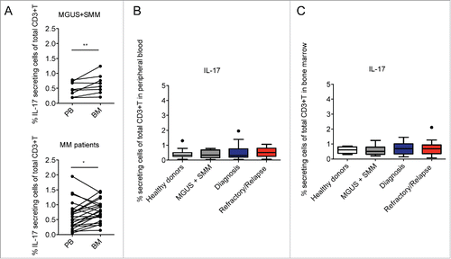
Figure 1. Percentage of Th17 cells in healthy donors and asymptomatic and symptomatic MM patients. (A) Analysis of IL-17+ cells of total CD3+ cells in paired samples of PB and BM mononuclear cells. Significant values calculated with the Wilcoxon matched-pairs signed rank test were indicated as: *p < 0.05 and **0.001< p <0.01. (B) Percentage of IL-17+ cells of total CD3+ cells in PB in different categories of subjects: Healthy donors (n = 15), MGUS+SMM (n = 11), Diagnosis (n = 20), Refractory/Relapse (n = 9). (C) Percentage of IL-17+ cells of total CD3+ cells in BM in different categories of subjects: Healthy donors (n = 4); MGUS+SMM (n = 10), Diagnosis (n = 19), Refractory/Relapse (n = 14). (B–C) No significant differences were observed between the different categories of subjects tested.

Figure 2. Percentage of Th22 cells and Th17 cells in relation to the clinical status and the presence or the absence of bone disease. Tukey plots of cumulative results from intracellular staining analyses of IL-22, IL-17 and IL-13 expression by CD3+ T cells in BM aspirates. (A–B) Percentage of Th17 (i.e., IL-17+) cells (A) and Th22 (i.e., IL-22+IL-17IL-13+) cells (B) in the BM of MM patients classified according to clinical status as: ISS Stage I+II (n = 12), Stage III (n = 7) and relapsed/refractory (Rel/Ref) (n = 14). (C–D) Percentage of Th17 (C) and Th22 (D) cells in the BM of MM patients grouped according to absence (N) (n = 11) or presence (Y) (n = 18) of bone disease. Responses significantly different by Mann Whitney U test are indicated as: *p < 0.05 and **0.001 < p < 0.01; ns: not significant.
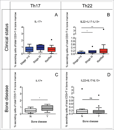
Figure 2. Percentage of Th22 cells and Th17 cells in relation to the clinical status and the presence or the absence of bone disease. Tukey plots of cumulative results from intracellular staining analyses of IL-22, IL-17 and IL-13 expression by CD3+ T cells in BM aspirates. (A–B) Percentage of Th17 (i.e., IL-17+) cells (A) and Th22 (i.e., IL-22+IL-17−IL-13+) cells (B) in the BM of MM patients classified according to clinical status as: ISS Stage I+II (n = 12), Stage III (n = 7) and relapsed/refractory (Rel/Ref) (n = 14). (C–D) Percentage of Th17 (C) and Th22 (D) cells in the BM of MM patients grouped according to absence (N) (n = 11) or presence (Y) (n = 18) of bone disease. Responses significantly different by Mann Whitney U test are indicated as: *p < 0.05 and **0.001 < p < 0.01; ns: not significant.

Reprints and Corporate Permissions

Please note: Selecting permissions does not provide access to the full text of the article, please see our help page How do I view content?

To request a reprint or corporate permissions for this article, please click on the relevant link below:

Academic Permissions

Please note: Selecting permissions does not provide access to the full text of the article, please see our help page How do I view content?

Obtain permissions instantly via Rightslink by clicking on the button below:

If you are unable to obtain permissions via Rightslink, please complete and submit this Permissions form. For more information, please visit our Permissions help page.