Figures & data
Figure 1. Kaplan–Meier survival analysis, macrophage infiltrates and phenotypes. Survival curves of patients with primary CRC in correlation with (A) tumor budding, (B) iCD68 and (C) sCD68 counts. (D) Survival of CRC patients in correlation with the frequency of cell-to-cell contacts between tumor buds and CD68+ macrophages in the tumor microenvironment. Survival curves of CRC patients with primary CRC dependent on (E) CD163+ and (F) iNOS+ macrophage counts.
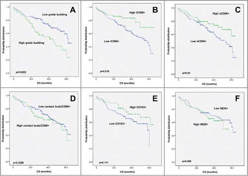
Figure 2. Representative images of macrophage infiltrates in the colorectal microenvironment (A) Intraepithelial and (B) stromal CD68+ macrophages (x100 each) (C) Direct cell-to-cell contacts between CD68+ macrophages and cytokeratin-positive tumor buds (x400) (D) Engulfment of cancer cells, as seen with pan-cytokeratin and CD68+ double immunohistochemistry (x400) (E) CD163+ M2 macrophage infiltrates (x100) (F) iNOS (brown) / MCT (red) double stain highlighting iNOS+/MCT− M1 macrophages (x100) (G) iNOS (brown) / CD163 (red) double stain showing co-existence of M1- and M2- polarized macrophages (x300) (H) Representative images from a tumor with a CD163+ (red) dominant M2-macrophage infiltrate and (I) from a tumor with an iNOS+ (brown) M1-macrophage infiltrate (CD163/iNOS double stain; x300).
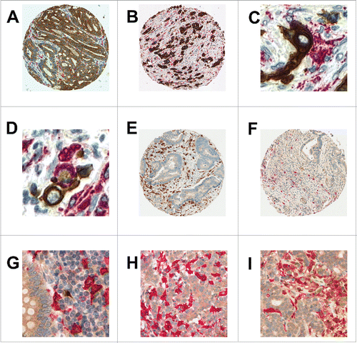
Table 1. Association of intratumoral (i) and stromal (s) CD68+ cells (normalized by stroma per punch) with clinicopathological and molecular features of CRC (n = 201).
Table 2. Multivariable Cox regression analysis of iCD68 and sCD68 counts in CRC adjusting for pT, pN, pM and post-operative therapy.
Table 3. Association of CD68+ buds contact, stromal CD163 and iNOS (normalized by percent stroma per TMA spot) with clinicopathological features.
Table 4. Association of CD47 expression with clinicopathological and molecular features (n = 182).
Figure 3. Kaplan–Meier survival analysis, CD47 expression. (A) Survival curves of patients with primary CRC in correlation with high or low CD47 expression. (B) Subgroup analysis of CD47 expression in tumors with strong CD163+ macrophage infiltration. (C) Subgroup analysis of patients with CD47 negative tumors and strong macrophage infiltration in comparison to all other marker combinations.
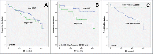
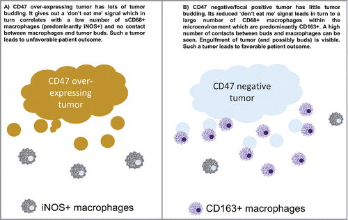
Figure 4. Working model.