Figures & data
Figure 1. The production of IFNγ by CAR19 T cells following recognition of B-cell lymphoma cell line Ramos is enhanced by Lenalidomide. CAR19 T cells were incubated at 10:3 ratio with Ramos B-cells overnight, in the presence or absence of 10 MM lenalidomide and the production of IFNγ into culture supernatant was determined with ELISA. The experiment was performed twice with similar results.
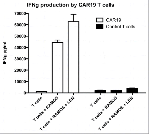
Figure 2. Lenalidomide enhances signaling via CAR19 receptor. To determine the costimulatory effect of lenalidomide, CAR19 T cells were stimulated with immobilized anti-CD3 antibody (clone MEM-57) or, with immobilized anti-CAR serum (polyclonal goat anti-mouse IgG FAB2) in the presence (10 MM, 1 MM) or absence of lenalidomide. The production of IFNγ into culture supernatant was determined with ELISA after overnight co-incubation. The experiment was performed twice with similar results.
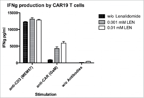
Figure 3. Lenalidomide acts directly onto T cells but not B cells to costimulate signaling via CAR19. (A) The production of IFNγ was measured by ELISA following pretreatment of CAR19 T cells or RAMOS cells for 16 h with 10 MM LEN, after wash-out of excess LEN cells were further co-incubated at 10:3 ratio overnight and production of IFNγ was then determined in culture supernatant. (B) The upregulation of activatory marker CD69 following recognition of Ramos B cells was determined on CAR+ T cells by flow cytometry in same setup as in , T cells were labeled with CFSE to easily separate them from B cells, the expression of CAR19 was visualized with Alexa 647 labeled goat anti-mouse serum. Combinations of either pretreated CAR19 T cells (CAR+L, Ramos-L), or Ramos B cells (CAR-L, Ramos+L), or cells without LEN (CAR-L, Ramos-L) are shown. The expression of CD69 shown in histograms was gated on either CAR19 T cells expressing high amounts, low amounts or negative for CAR19. These experiments were performed twice with similar results.
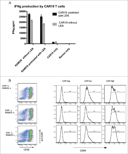
Figure 4. Analysis of changes in protein expression induced by LEN. (A) Downregulation of Ikaros and Aiolos by LEN. CAR19 T cells were pretreated with LEN 10 MM for 24 h (+), or without it (−), cells were then lysed in SDS loading buffer and the levels of Ikaros and Aiolos transcription factors were determined by immunoblotting together with the ubiquitin ligase Cereblon, actin indicates the equal loading. (B) Enhanced ERK activation by ligation of CAR after LEN pre-treatment (10 MM for 24 h). CAR19 T cells were activated by immobilized anti-CAR antibody (goat anti-mouse), or by anti-CD3 antibody (clone MEM-57) for 30 min at 37°C, cells were then lysed in SDS sample buffer and the phosphorylation status of ERK was determined by immunoblotting, the total levels of ERK are shown in the bottom panel. Both experiments were performed twice with similar results.
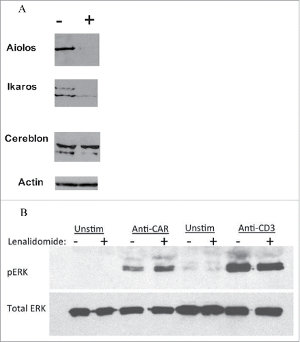
Figure 5. LEN enhances antitumor response in vivo to TMD8 DLBCL cells. NSG mice were transplanted SCl with 5 million DLBCL ABC cells TMD8 and then received two doses of 5 million. CAR20 T cells followed with daily IP injection of 10 Mg of LEN, 14 d later mice were sacrificed and the tumors were excised and weighted (A). In panel B we shown the infiltration of tumors by CD8+ T cells which was determined by flow cytometry in cell suspension prepared from excised tumors. This experiment was performed twice with similar results.
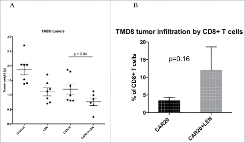
Figure 6. LEN enhances antitumor response in vivo to Ramos Burkitt lymphoma cells. NSG mice were transplanted SC with 5 million Ramos cells and then received one dose of 5 million CAR19 T cells followed with daily IP injection of LEN, 21 d later mice were sacrificed, the tumors were excised and weighted (A). In panel B is shown the infiltration of tumors by CD8+ T cells which was determined by flow cytometry in cell suspension prepared from excised tumors. This experiment was performed once.
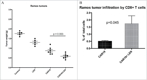
Figure 8. LEN enhances response antitumor response in vivo to established primary diffuse large B cell lymphoma (A) NSG mice were SC transplanted with 10 million DLBC cell KTC followed with one doses of 5 million CAR19 T cells and daily IP injection LEN, 14 d later mice were sacrificed, the tumors were excised and weighted. (B) NSG mice were SC transplanted with 5 million DLBC cell KTC and left for 10 d to establish cca 0.5 cm tumors, during this period all mice were receiving daily injections LEN. After the tumors were established, mice received three doses of 5 million. CAR19 T cells at days 10, 12 and 17 with, or without LEN. Two weeks after treatment with CAR19 T cells, mice were sacrificed and the size of tumors was determined. The experiment in was performed once.
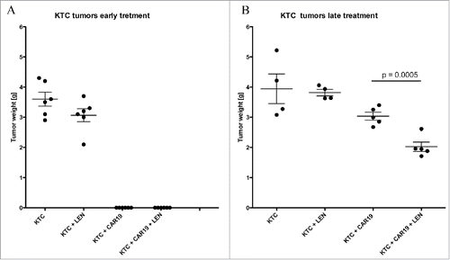
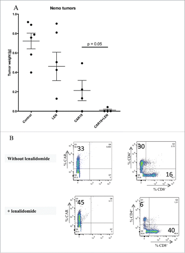
Figure 7. Lenalidomide enhances antitumor response in vivo to primary mantle cell lymphoma. (A) NSG mice received SC 5 million. NEMO cells followed by IV injection of two doses of 5 million CAR19 T cells and daily IP injection of LEN, 14 d later mice were sacrificed and the tumors were excised weighted. (B) The infiltration of tumors by T cells was analyzed by flow cytometry in a cell suspension prepared from excised tumors using antibodies to CD4+, CD8+ and CAR, one representative mouse is shown. The experiment in was performed once.