2,002
Views
34
CrossRef citations to date
0
Altmetric
Original Research

ADAM10 new selective inhibitors reduce NKG2D ligand release sensitizing Hodgkin lymphoma cells to NKG2D-mediated killing

, , , , , , , , & show all
Article: e1123367 | Received 07 Aug 2015, Accepted 18 Nov 2015, Published online: 11 May 2016

Figures & data

Figure 1. The mature membrane form of ADAM10 is expressed on HL cells. Panels (A), (B), (D) Lysates obtained from HL LN cell suspensions (A) or HL cell lines (B) or LN MSCs obtained by culturing LN cell suspensions from HL patients (D) were subjected to Western blot as described in Materials and Methods; membranes were probed with the anti-ADAM10 or anti-β actin mAb followed by the relevant HRP-conjugated secondary antibodies and developed with the HRP substrate. In each blot, the precursor form (p) and the mature form (m) of ADAM10 molecule is indicated. Panels (C) and (E) Surface expression of ADAM10 on KMH2, L540, L428 (Ca, Cb, Cc, dark gray histograms) or MSC773 or RS773 (Ea, Eb) was evaluated with the specific mAb directed against the mature form of ADAM10 followed by APC-conjugated GAM and FACS analysis; results are expressed as Log far red fluorescence intensity, a.u., vs. number of cells.
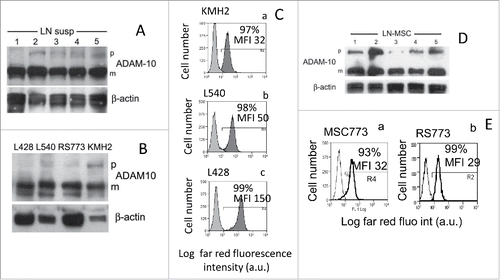
Figure 1. The mature membrane form of ADAM10 is expressed on HL cells. Panels (A), (B), (D) Lysates obtained from HL LN cell suspensions (A) or HL cell lines (B) or LN MSCs obtained by culturing LN cell suspensions from HL patients (D) were subjected to Western blot as described in Materials and Methods; membranes were probed with the anti-ADAM10 or anti-β actin mAb followed by the relevant HRP-conjugated secondary antibodies and developed with the HRP substrate. In each blot, the precursor form (p) and the mature form (m) of ADAM10 molecule is indicated. Panels (C) and (E) Surface expression of ADAM10 on KMH2, L540, L428 (Ca, Cb, Cc, dark gray histograms) or MSC773 or RS773 (Ea, Eb) was evaluated with the specific mAb directed against the mature form of ADAM10 followed by APC-conjugated GAM and FACS analysis; results are expressed as Log far red fluorescence intensity, a.u., vs. number of cells.

Figure 2. ADAM10 silencing leads to decreased shedding of NKG2D-L. KMH2 and L428 cells were transfected with ADAM10 (siADAM10) or ADAM17 (siADAM17) siRNA or non-targeting siRNA (siNT) pool as negative control (KMH2: panels A, B; L428: panels C, D). Protein expression was analyzed by Western blot (panels Aa and Ca), and FACS analysis (Ab and Cb; in each histogram the percentage and MFI of positive cells is shown) with the specific anti-ADAM10 or anti-ADAM17 antibodies, 72 h after transfection. Soluble MIC-B (Ba, Da) or ULBP3 (Bb, Db) or sALCAM (Bc, Dc) were evaluated by ELISA in SN (collected upon further 24 h of culture 72 h after transfection). Results in (B) and (D) are expressed as pg/mL/105 cells and are the mean ± SD from three independent experiments. *p <0.001 vs siNT.
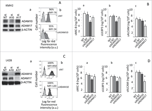
Figure 2. ADAM10 silencing leads to decreased shedding of NKG2D-L. KMH2 and L428 cells were transfected with ADAM10 (siADAM10) or ADAM17 (siADAM17) siRNA or non-targeting siRNA (siNT) pool as negative control (KMH2: panels A, B; L428: panels C, D). Protein expression was analyzed by Western blot (panels Aa and Ca), and FACS analysis (Ab and Cb; in each histogram the percentage and MFI of positive cells is shown) with the specific anti-ADAM10 or anti-ADAM17 antibodies, 72 h after transfection. Soluble MIC-B (Ba, Da) or ULBP3 (Bb, Db) or sALCAM (Bc, Dc) were evaluated by ELISA in SN (collected upon further 24 h of culture 72 h after transfection). Results in (B) and (D) are expressed as pg/mL/105 cells and are the mean ± SD from three independent experiments. *p <0.001 vs siNT.

Figure 3. ADAM10 inhibitors reduce the shedding of NKG2D-L by HL cell lines and maintain the binding of NKG2D receptor. L428 cells were exposed to culture medium alone, DMSO or GI254023X (GIX), JG26, MN8 or LT4 (at 10 to 2.5 μM concentration) for 24 h (panel A), followed by 100 μM Na3VO4 as pervanadate for 40 min at 37°C (panel B and C). Then, SN were harvested and sMIC-A (Aa, Ba), sMIC-B (Ab, Bb), sULBP2 (Ac, Bc), sULBP3 (Ad, Bd) or sALCAM (C) measured by specific ELISA. Results are expressed as ng/mL/106 cells and are representative of four independent experiments. *p <0.001 vs. DMSO. Panels (D) and (E) L428 cells exposed for 24 h to DMSO or 10 μM LT4 or 100 μM Na3VO4 as pervanadate, in the absence or presence of 10 μM LT4 as indicated, were harvested and evaluated for the expression of ULBP2 (D) with the specific mAb followed by APC-conjugated GAM or for the binding of the chimeric receptor (FcNKG2D, panel E) followed by APC-conjugated anti-human Fc antiserum, by FACS analysis; results are expressed as Log far red fluorescence intensity (arbitrary units, a.u.) vs. number of cells. In each subpanel: percentage and mean fluorescence intensity (MFI, a.u.) of positive cells. One representative experiment out of four.
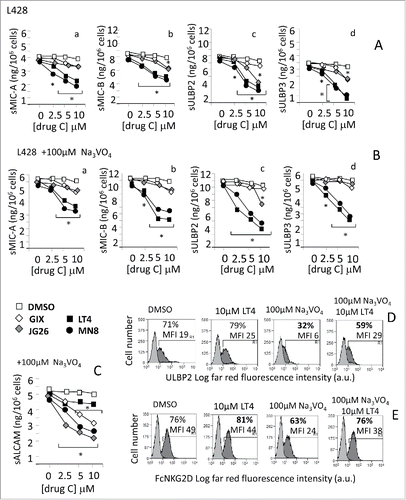
Figure 3. ADAM10 inhibitors reduce the shedding of NKG2D-L by HL cell lines and maintain the binding of NKG2D receptor. L428 cells were exposed to culture medium alone, DMSO or GI254023X (GIX), JG26, MN8 or LT4 (at 10 to 2.5 μM concentration) for 24 h (panel A), followed by 100 μM Na3VO4 as pervanadate for 40 min at 37°C (panel B and C). Then, SN were harvested and sMIC-A (Aa, Ba), sMIC-B (Ab, Bb), sULBP2 (Ac, Bc), sULBP3 (Ad, Bd) or sALCAM (C) measured by specific ELISA. Results are expressed as ng/mL/106 cells and are representative of four independent experiments. *p <0.001 vs. DMSO. Panels (D) and (E) L428 cells exposed for 24 h to DMSO or 10 μM LT4 or 100 μM Na3VO4 as pervanadate, in the absence or presence of 10 μM LT4 as indicated, were harvested and evaluated for the expression of ULBP2 (D) with the specific mAb followed by APC-conjugated GAM or for the binding of the chimeric receptor (FcNKG2D, panel E) followed by APC-conjugated anti-human Fc antiserum, by FACS analysis; results are expressed as Log far red fluorescence intensity (arbitrary units, a.u.) vs. number of cells. In each subpanel: percentage and mean fluorescence intensity (MFI, a.u.) of positive cells. One representative experiment out of four.

Table 1. In vitro enzymatic activity (IC50nM values)a of new compounds LT4 and MN8 and the reference compounds JG26 and GI254023X.

Figure 4. Exposure to ADAM10 inhibitors increases the sensitivity of HL cell lines to NKG2D-dependent cell killing. Cytolytic activity of NK cells (n = 6, panel A and B) or γδ T cells (n = 6, panel C) was analyzed against L428 (Aa, Ab, Ca) or L540 (Ba, Bb, Cb) cell lines at E:T ratio of 10:1 in a 4-h 51Cr-release assay. Some samples were set up after exposure of the target cell lines to either medium, or DMSO or LT4 or MN8 (Aa, Ba, Ca, Cb), GIX or JG26 (Ab, Bb) at 10 μM concentration for 24 h. In some samples, effector cells were exposed to saturating amounts (5 μg/mL) of the anti-NKG2D mAb at the onset of the cytotoxicity assay; an unrelated mAb, matched for the isotype, used as control, did not exert any effect (not shown). Results are expressed as % specific lysis calculated as described in Materials and Methods.
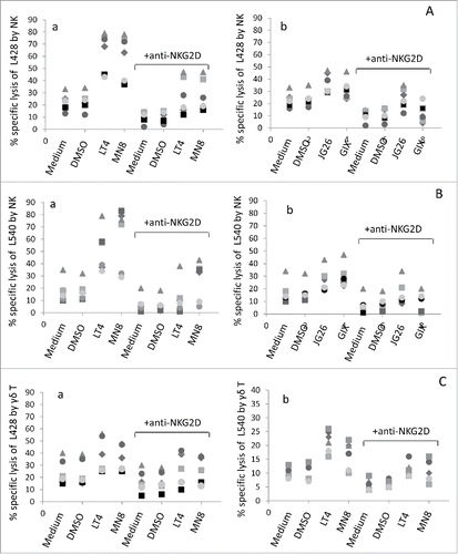
Figure 4. Exposure to ADAM10 inhibitors increases the sensitivity of HL cell lines to NKG2D-dependent cell killing. Cytolytic activity of NK cells (n = 6, panel A and B) or γδ T cells (n = 6, panel C) was analyzed against L428 (Aa, Ab, Ca) or L540 (Ba, Bb, Cb) cell lines at E:T ratio of 10:1 in a 4-h 51Cr-release assay. Some samples were set up after exposure of the target cell lines to either medium, or DMSO or LT4 or MN8 (Aa, Ba, Ca, Cb), GIX or JG26 (Ab, Bb) at 10 μM concentration for 24 h. In some samples, effector cells were exposed to saturating amounts (5 μg/mL) of the anti-NKG2D mAb at the onset of the cytotoxicity assay; an unrelated mAb, matched for the isotype, used as control, did not exert any effect (not shown). Results are expressed as % specific lysis calculated as described in Materials and Methods.

Figure 5. Improvement of HL cell lysis by exposure to ADAM10 inhibitor LT4 and anti-TGFβ. Panel (A) NKG2D expression before (upper histograms) or after treatment with TGFβ (10 ng/mL), (middle histograms) or with TGFβ and anti-TGFβ mAb (1 µg/mL), on NK cells (Aa) or γδ T cells (Ab). In each subpanel: percentage of positive cells and MFI (a.u.). Panel (B) Cytolytic activity of NK cells (Ba) orγδ T cells (Bb) was analyzed against L428 cell line at E:T ratio of 5:1 in a 4-h 51Cr-release assay. Some samples were set up after exposure of the target cell lines to LT4 or MN8 at 10 μM concentration for 24 h. To some samples, we added effector cells exposed to TGFβ (10 ng/mL), with or without saturating amounts (1 μg/mL) of the anti-TGFβ mAb, as indicated. Results are expressed as % inhibition or stimulation of specific lysis calculated as described in Materials and Methods. *p <0.001 vs. TGFβ. **p <0.001 vs. TGFβ + anti-TGFβ. #p <0.001 vs. TGFβ + anti-TGFβ on untreated L428 cells.
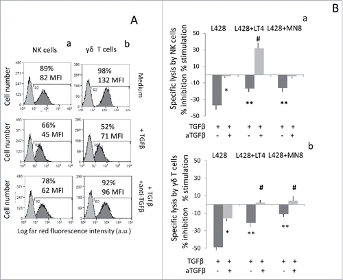
Figure 5. Improvement of HL cell lysis by exposure to ADAM10 inhibitor LT4 and anti-TGFβ. Panel (A) NKG2D expression before (upper histograms) or after treatment with TGFβ (10 ng/mL), (middle histograms) or with TGFβ and anti-TGFβ mAb (1 µg/mL), on NK cells (Aa) or γδ T cells (Ab). In each subpanel: percentage of positive cells and MFI (a.u.). Panel (B) Cytolytic activity of NK cells (Ba) orγδ T cells (Bb) was analyzed against L428 cell line at E:T ratio of 5:1 in a 4-h 51Cr-release assay. Some samples were set up after exposure of the target cell lines to LT4 or MN8 at 10 μM concentration for 24 h. To some samples, we added effector cells exposed to TGFβ (10 ng/mL), with or without saturating amounts (1 μg/mL) of the anti-TGFβ mAb, as indicated. Results are expressed as % inhibition or stimulation of specific lysis calculated as described in Materials and Methods. *p <0.001 vs. TGFβ. **p <0.001 vs. TGFβ + anti-TGFβ. #p <0.001 vs. TGFβ + anti-TGFβ on untreated L428 cells.
Supplemental material

KONI_A_1123367_supplemental_material.zip

Download Zip (852.3 KB)