Figures & data
Table 1. Clinicopathological data of the 87 breast cancer patients investigated. Clinicopathological data and group distribution of the 87 breast cancer patients with respect to the four anatomical locations at which the corresponding metastasis had occurred. Percentages are shown in relation to the respective metastatic site.
Figure 1. Distribution pattern of TIL subsets in the three tumor compartment of a representative primary breast cancer and its corresponding brain metastasis. Illustration of a primary breast carcinoma and its corresponding brain metastasis with HE as well as CD4+, CD8+ and CD20 immunostainings in all three examined tumor compartments are depicted (iTIL: intratumoral infiltrating lymphocytes, sTIL: stromal infiltrating lymphocytes, imTIL: infiltrative margin infiltrating lymphocytes).
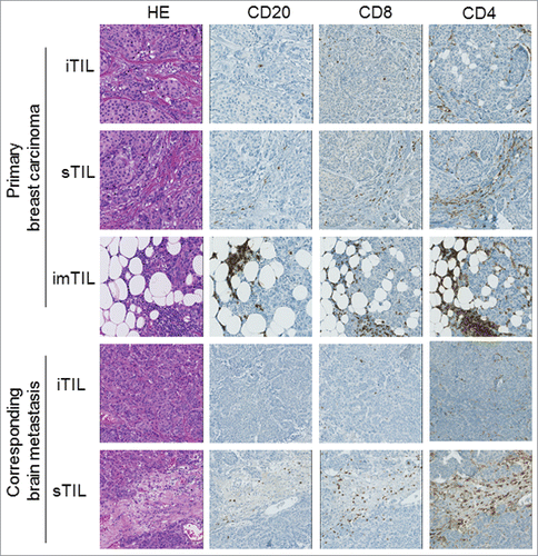
Figure 2. Similar distribution of TIL with regard to the investigated primary tumor compartments and the lymphocyte subtypes but irrespectively of the site of distant metastasis. Scatter graphs show that within the primary tumor compartments (iTIL (not shown), sTIL and imTIL) invasive margin TIL (imTIL) are significantly increased when compared to stromal (sTIL) TIL irrespectively of the particular site (A–D) to which metastasis had occurred. For all three compartments (iTIL not shown) CD4+, CD8+ and CD20+ lymphocyte subsets always followed the same gradient with CD4+ lymphocytes being the most prominent, followed by CD8+ lymphocytes and at last CD20+ lymphocytes again irrespectively of the anatomical site of distant metastasis (A–D). Significances are displayed as follows: ns = p > 0.05, *p ≤ 0.05, **p ≤ 0.01, ***p ≤ 0.001 and ****p ≤ 0.0001.
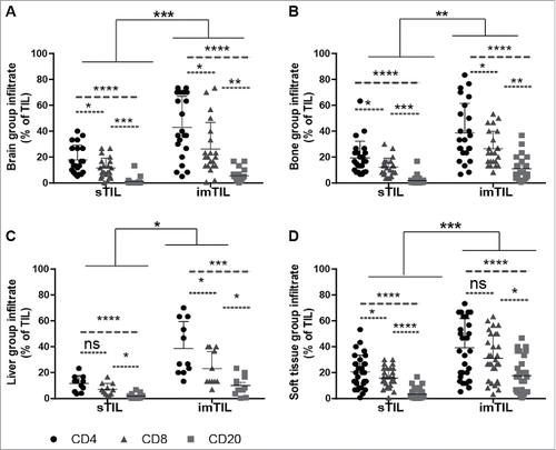
Table 2. Distribution of tumor infiltrating lymphocytes (TIL) subsets within the primary tumor. Mean and median are displayed for CD4+, CD8+ and CD20+ lymphocytes within the three investigated primary tumor compartments consisting of intratumoral (iTIL), stromal (sTIL) and invasive margin (imTIL) TIL divided into the four anatomical sites (brain, bone, liver and soft tissue) of the corresponding metastasis.
Table 3. Distribution of metastasis infiltrating lymphocytes (mTIL) subsets within the site of metastasis. Mean and median are displayed for CD4+, CD8+ and CD20+ lymphocytes within the two investigated metastasis compartments consisting of intratumoral (iTIL) and stromal (sTIL) divided into the four anatomical sites (brain, bone, liver and soft tissue) of the corresponding metastasis.
Figure 3. Significantly more sTIL within the primary tumor than mTIL within the site of the corresponding metastasis. Bar graphs for CD4+, CD8+ and CD20+ stromal infiltrating lymphocyte subsets show that there are significantly more sTIL within the primary tumor than mTIL within the site of the corresponding metastasis irrespectively of the anatomical site of distant metastasis (A–D). The distribution of infiltrating lymphocytes is schematically visualized with respect to the three primary tumor/metastasis compartments (E) consisting of intratumoral lymphocytes (iTIL_PT/iTIL_Met = white dots), stromal lymphocytes (sTIL_PT/sTIL_Met = gray dots) and invasive margin lymphocytes (imTIL_PT/imTIL_Met = black dots) and with respect to the lymphocyte subsets within these compartments (F) comprising CD4+ infiltrating lymphocytes (red dots), CD8+ infiltrating lymphocytes (green dots) and CD20+ infiltrating lymphocytes (blue dots). Significances are displayed as follows: ns = p > 0.05, *p ≤ 0.05, **p ≤ 0.01, ***p ≤ 0.001 and ****p ≤ 0.0001.
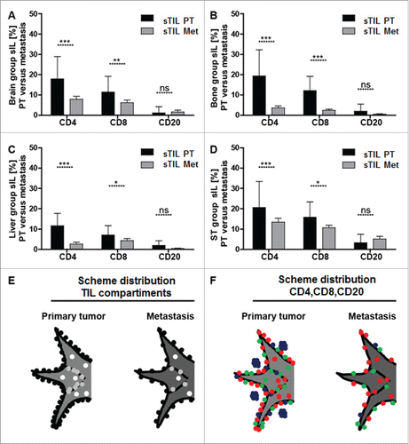
Figure 4. Higher amounts of CD8+ lymphocytes within the invasive margin is associated with significantly longer disease free survival (DFS). Kaplan–Meier plots of disease free survival dichotomized at the respective CD8+ imTIL median of the primary tumor (also see ) into CD8+ T cells high and CD8+ T cells low show that higher amounts of CD8+ T cells within the invasive margin of the primary tumor are generally (A) but also in the subgroups with distant metastasis into brain and bone except for soft tissue metastasis associated with a with significantly longer DFS again (B–D).
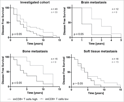
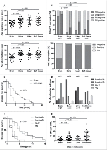
Figure 5. Clinico-pathological correlation of intrinsic subtypes and different metastastic sites of breast cancer to overall survival and to CD20 distribution at the invasion front. Brain metastasis group shows different clinicopathological and CD20+ imTIL behavior. Patients with distant metastasis to the brain show a significantly younger age at onset of both primary tumor and metastasis when compared to the other groups (A). Time to occurrence of distant brain metastasis (= disease free survival) is significantly reduced in the brain group when compared to all other groups (B). Patients with brain metastasis show significantly less estrogen receptor (ER) and progesterone (PR) but significantly more Her2 receptor expression in the primary tumor (C,D). Curiously, CD20 tumor infiltrating lymphocytes at the infiltrative margin (imTIL) of the primary tumor are significantly reduced in the brain group when compared to the other groups where distant metastasis had occurred to bone, liver or soft tissue (E). When correlating intrinsic subtypes to overall survival, Luminal A and Luminal B tumors are separated from HER2 positive and TN breast cancer cases by a similarly improved overall survival (F).
