Figures & data
Figure 1. Myeloid cell infiltration into murine pancreatic tissue. Immunhistochemical staining for CD68 in representative pancreatic tissues of LSL-KrasG12D/+; LSL-Trp53R172H/+; Pdx-1-Cre mice. (A) Normal area. (B) PanIN lesion. (C) Invasive PDAC. Bars indicate 100 μm.
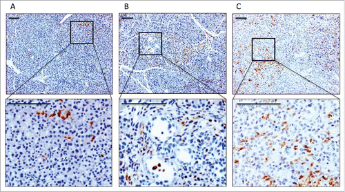
Figure 2. Myeloid cells show an increase in M2 marker gene expression during pancreatic carcinogenesis. qRT-PCR analysis of the mRNA levels of the M2 marker genes MSR-1 (A) and MRC-1 (B) in CD11b+ cells isolated from pancreata of WT, KC and KPC mice. (C, D) Protein expression of MSR-1 (C), MRC-1 (D) as detected by FACS. *p < 0.05, **p < 0.01.
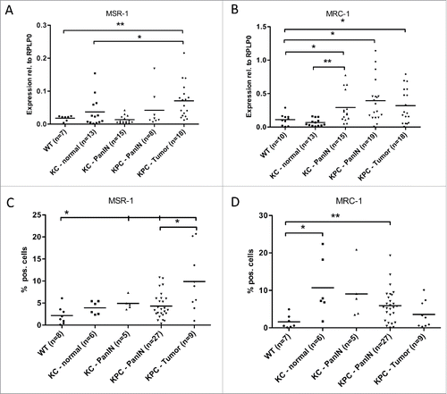
Figure 3. Myeloid cells show an increased M1 marker gene expression during pancreatic carcinogenesis. qRT-PCR analysis of the mRNA levels of the M1 marker genes CCR-7 (A) and MHC-2 (B) in CD11b+ cells isolated from pancreata of WT, KC and KPC mice. (C, D) Protein expression of CCR-7 (C) and MHC-2 (D) as detected by FACS. *p < 0.05, **p < 0.01.
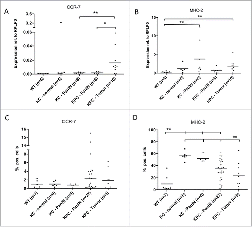
Figure 4. Expression of miR-21-3p and miR-21-5p increases during pancreatic carcinogenesis. (A) qRT-PCR Profiler for miRNAs in CD11b+ cells isolated from wild-type mice and KPC-mice with invasive PDAC. Green dots represent housekeeping genes. B+C, qRT-PCR analysis of miR-21-3p (B) and miR-21-5p (C) levels in CD11b+ cells isolated from pancreata of WT, KC and KPC mice. *p < 0.05, *p < 0.01.
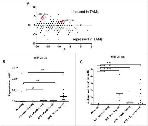
Figure 5. miR-21 inhibits LPS-induced expression of CCL-3/MIP-1α and CXCL-10/IP-10. (A, B) CCL-3/MIP-1α protein levels determined by ELISA in cell culture supernatants of murine bone marrow-derived macrophages +/− LPS (10 ng/mL) and +/− mimics for miR-21-3p (A) and miR21-5p (B). (C, D) CXCL-10 protein levels determined by ELISA in the cell culture supernatants of murine bone marrow-derived macrophages +/− LPS (10 ng/mL) and +/− mimics for miR-21-3p (C) and miR21-5p (D). *p < 0.05.
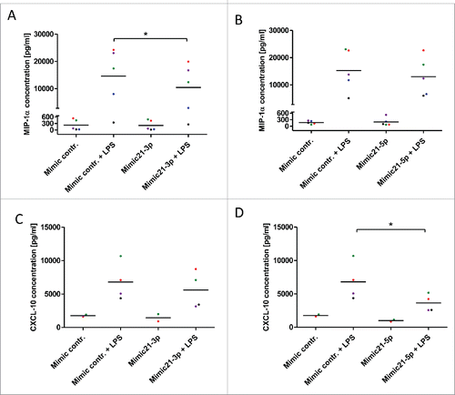
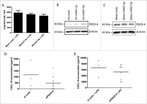
Figure 6. miR-21 mediates an immunosuppressive phenotype. (A) CD8+ T-cells isolated from murine spleen and lymph nodes were co-cultured in a transwell assay with conditioned media of murine bone marrow-derived macrophages stimulated with LPS (10 ng/mL) +/− mimics for miR-21-3p or +/− mimics for miR-21-5p. (B, C) Immunoblot analysis of PDCD-4 in murine bone marrow-derived macrophages +/− inhibitor miR-21-5p (B) and +/− mimic miR-21-5p (C). (D, E) CXCL-10 protein levels determined by ELISA in cell culture supernatants of murine bone marrow-derived macrophages +/− LPS (10 ng/mL) and +/− PDCD-4 siRNA. *p < 0.05.