870
Views
16
CrossRef citations to date
0
Altmetric
Brief Report

CD137 signaling in Hodgkin and Reed-Sternberg cell lines induces IL-13 secretion, immune deviation and enhanced growth

, &
Article: e1160188 | Received 16 Dec 2015, Accepted 25 Feb 2016, Published online: 31 May 2016

Figures & data

Figure 1. CD137 signaling induces IL-13, TNF and IL-6 secretion by HRS cell lines. 5 × 10⁵ L-428-control, L-1236-control, L-428-CD137, L-1236-CD137 or KM-H2 cells were cultured on plates coated with 5 μg/mL of recombinant CD137L or BSA for 24 h. Levels of IL-13 (A), TNF (B) and IL-6 (C) were measured by ELISA. Depicted are means ± SD of triplicate measurements. *p <0.05; **p <0.01; ***p <0.005. Data are representative of three independent experiments.
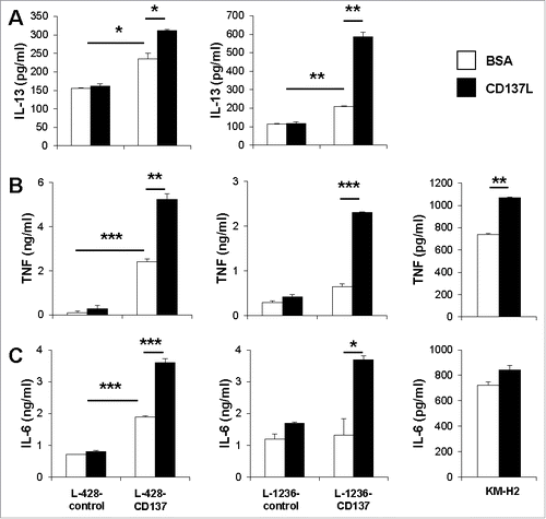
Figure 1. CD137 signaling induces IL-13, TNF and IL-6 secretion by HRS cell lines. 5 × 10⁵ L-428-control, L-1236-control, L-428-CD137, L-1236-CD137 or KM-H2 cells were cultured on plates coated with 5 μg/mL of recombinant CD137L or BSA for 24 h. Levels of IL-13 (A), TNF (B) and IL-6 (C) were measured by ELISA. Depicted are means ± SD of triplicate measurements. *p <0.05; **p <0.01; ***p <0.005. Data are representative of three independent experiments.
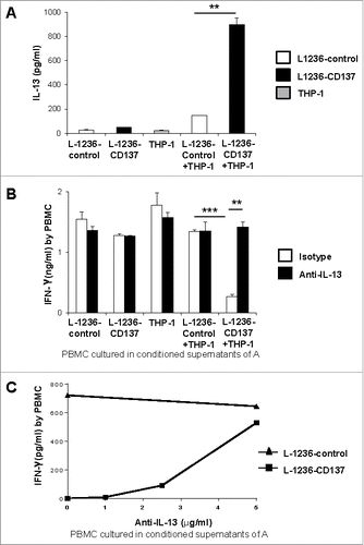
Figure 2. (A) 5 × 10⁵ L-1236-control or L-1236-CD137 cells were co-cultured with 5 × 10⁵ THP-1 cells for 24 h. Supernatants from the cocultures were tested for IL-13 levels by ELISA. (B) PBMC were sub-optimally activated with 2 ng/mL of anti-CD3 (clone OKT3) and cultured in 50% conditioned supernatants of the cocultures of (A) for 24 h. 5 μg/mL of polyclonal goat IgG or goat polyclonal IL-13 antibody were added. (C) Anti-IL-13 antibody has been added at indicated concentrations to activated PBMC cultured in 50% conditioned supernatants of (A). IFNγ secretion by was measured by ELISA after 24 h. Depicted are means ± SD of triplicate measurements. *p <0.05; **p <0.01; ***p <0.005. Data are representative of three independent experiments.

Figure 2. (A) 5 × 10⁵ L-1236-control or L-1236-CD137 cells were co-cultured with 5 × 10⁵ THP-1 cells for 24 h. Supernatants from the cocultures were tested for IL-13 levels by ELISA. (B) PBMC were sub-optimally activated with 2 ng/mL of anti-CD3 (clone OKT3) and cultured in 50% conditioned supernatants of the cocultures of (A) for 24 h. 5 μg/mL of polyclonal goat IgG or goat polyclonal IL-13 antibody were added. (C) Anti-IL-13 antibody has been added at indicated concentrations to activated PBMC cultured in 50% conditioned supernatants of (A). IFNγ secretion by was measured by ELISA after 24 h. Depicted are means ± SD of triplicate measurements. *p <0.05; **p <0.01; ***p <0.005. Data are representative of three independent experiments.

Figure 3. CD137 expression increases HRS cell proliferation. Indicated numbers of L-428 (A) or L-1236 (B) or KM-H2 (C) cells were seeded (white bar: non-CD137-expressing cells, black bar: CD137-expressing cells), and pulsed with 0.5 μCi of 3H-thymidine at the same time. The cells were harvested 24 h later, and 3H-thymidine incorporation was quantified. Depicted are means ± SD. *p <0.05, **p <0.01, ***p < 0.001.
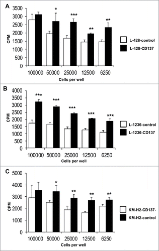
Figure 3. CD137 expression increases HRS cell proliferation. Indicated numbers of L-428 (A) or L-1236 (B) or KM-H2 (C) cells were seeded (white bar: non-CD137-expressing cells, black bar: CD137-expressing cells), and pulsed with 0.5 μCi of 3H-thymidine at the same time. The cells were harvested 24 h later, and 3H-thymidine incorporation was quantified. Depicted are means ± SD. *p <0.05, **p <0.01, ***p < 0.001.

Figure 5. Schematic representation of the role of CD137 in immune deviation and growth stimulation of HRS cells.
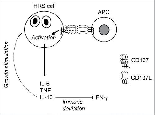
Figure 5. Schematic representation of the role of CD137 in immune deviation and growth stimulation of HRS cells.

Table 1. Expression of CD137 and IL-13 in HL tumor microarray determined by immunohistochemical double staining. For CD137: + at least 5 cells, ++ at least 10 cells, +++ more than 15 cells stained in the section. For IL-13 staining: + at least 10%, ++ at least 30% and +++ more than 50% of the section stained.

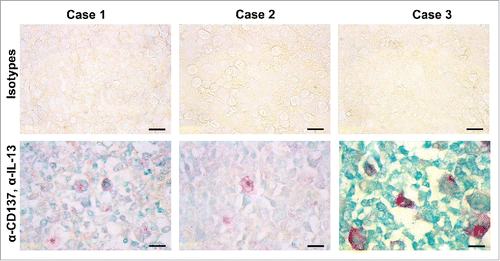
Figure 4. CD137-positive cells in HL express IL-13. Immunohistochemical detection of IL-13 (green) and CD137 (red). Shown are three cases of classical HL of the mixed cellularity type. Size bar: 10 μm.

Figure 4. CD137-positive cells in HL express IL-13. Immunohistochemical detection of IL-13 (green) and CD137 (red). Shown are three cases of classical HL of the mixed cellularity type. Size bar: 10 μm.
Supplemental material

KONI_A_1160188_s02.pptx

Download MS Power Point (160.1 KB)

Reprints and Corporate Permissions

Please note: Selecting permissions does not provide access to the full text of the article, please see our help page How do I view content?

To request a reprint or corporate permissions for this article, please click on the relevant link below:

Academic Permissions

Please note: Selecting permissions does not provide access to the full text of the article, please see our help page How do I view content?

Obtain permissions instantly via Rightslink by clicking on the button below:

If you are unable to obtain permissions via Rightslink, please complete and submit this Permissions form. For more information, please visit our Permissions help page.