Figures & data
Figure 1. HER-2 mimotope AAV model and immunogenicity evaluation. (A) Model of the AAV vector displaying HER-2 specific mimotopes fused to its capsid protein VP3. Mimotopes are inserted between amino acid positions 587 and 588, which resemble a peak in the capsid, for optimal display. (B) AAV vectors displaying HER-2 mimotopes are able to induce HER-2 specific antibodies. Screening of sera from mice, immunized with AAV-displaying HER-2 mimotopes and Al(OH)3 as adjuvant, against the extracellular domain of human HER-2. Depicted are means (n = 2/mouse serum) & SD; values of MIS4 were statistically analyzed (One-way ANOVA, Tukey's post-test). (C) Antibodies induced by AAV clone DMD4 recognize HER-2 on the surface of cancer cells.Immunofluorescence staining of D2F2/E2 cells, overexpressing human HER-2 with sera of mice immunized with AAV clone DMD4 (PIS = Pre-Immune Serum, MIS 3 = MIS after three immunizations). After immunization with the HER-2 specific mimotope, membrane specific staining can be seen. Pre-Immune sera and serum of mice immunized with wtAAV alone do not show any specific staining on D2F2/E2 cells.
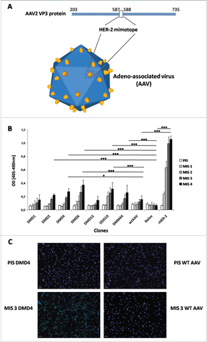
Table 1. Amino acid sequences of tested mimotopes.
Figure 2. Specificity and functionality testing of antibodies purified from sera of immunized mice. (A) AAV-mimotope induced antibodies recognize HER-2, but not tumor-associated antigens EGFR, CEA or control protein BSA. Antibodies (c = 1 µg/mL) purified from sera of naïve mice and mice immunized with candidate particles were screened for their reactivity toward known tumor antigens HER-2, EGFR, CEA as well as the control protein BSA in ELISA. Antibodies induced by mimotopes DMD4 and DMD6 show significant higher IgG reactivity toward rHER-2 than naive mice (p < 0.001) and mice immunized with candidates DMD1 (p < 0 .001) and DMD2 (p < 0 .001). Toward all other antigens only background reactivity is visible. Bars represent mean values & SD (n = 2); One-way ANOVA, Tukey's post-test. (B) Purified antibodies of immunized mice to different degrees inhibit growth of human HER-2 overexpressing mammary carcinoma cells. Tetrazolium-based proliferation assays with HER-2 overexpressing BT474 cells are depicted upon incubation for 72 h with purified antibodies (c = 1 µg/mL) of sera from mice immunized with candidate particles. Bars displaying mean values & SD, analyzed by means of Kruskal–Wallis test plus Dunns post-test.
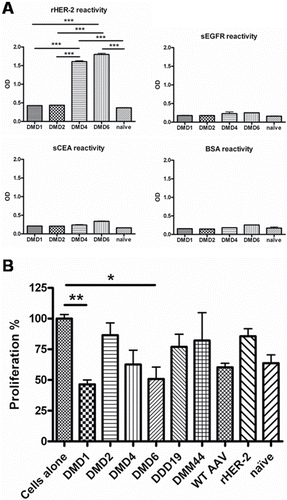
Figure 3. Subclass analysis of HER-2 specific antibodies induced by selected mimotope AAV clones DMD4 and DMD6 by ELISA. Left Panel: a-e: IgA, IgG1, IgG2a, IgG2b or IgE antibody determination before (PIS) and after each immunization (MIS 1-4). Right panel: Antibody levels after four immunization rounds (MIS 4); Box and whiskers plot, displaying minimum and maximum values. Each serum was measured in duplicate, eight mice per group ± SD; Kruskal–Wallis test, Dunns post-test.
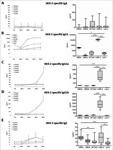
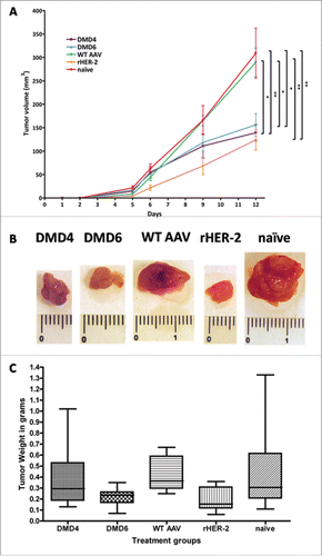
Figure 4. Readout of tumor graft trial. (A) Mice immunized with either rHER-2, DMD4 or DMD6 particles display significantly slower and smaller tumor growth compared to naive mice and mice immunized with wtAAV only. Tumor growth curve of grafted HER-2 overexpressing D2F2/E2 cells; at day 12, DMD4 immunized mice had significantly smaller tumors compared to those of the naive (p < 0.01) and of the wtAAV group (p < 0.05). DMD6 treated mice also developed significantly smaller tumors (p < 0.05 compared to naive group, p < 0.05 compared to wtAAV). Mice immunized with rHER-2 also developed significantly smaller tumors compared to both wtAAV treated (p < 0.01) or naive mice (p < 0.01). Tumor size at day 12 was analyzed by One-way ANOVA alongside Tukey's post-test; displayed are mean tumor volume values ± standard error of the mean (SEM). (B) Representative macroscopic pictures of explanted tumors. (C) Tumors of mice immunized with mimotope DMD6 or rHER-2 had significantly lower weight compared to tumors of animals receiving wtAAV. Diagram displaying weight of explanted tumors in grams; Box and whiskers plot, whiskers displaying minimum and maximum values (eight mice/group). Analysis by means of Kruskal–Wallis test and Dunns post-test.