1,443
Views
27
CrossRef citations to date
0
Altmetric
Review

Embracing rejection: Immunologic trends in brain metastasis

, , , , , , , & show all
Article: e1172153 | Received 18 Dec 2015, Accepted 23 Mar 2016, Published online: 29 Jun 2016

Figures & data

Figure 1. Tumor seeding and progression. The critical steps required for successful metastasis formation within the brain include (1) vascular arrest by size exclusion, (2) active extravasation of metastatic tumor cells, (3) stringent perivascular location and proliferation, and (4) sustained growth via angiogenesis and vascular remodeling.
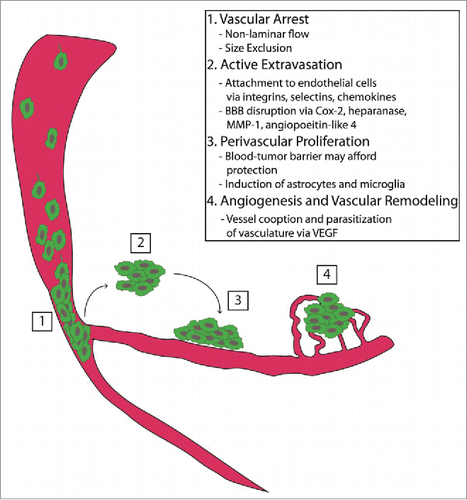
Figure 1. Tumor seeding and progression. The critical steps required for successful metastasis formation within the brain include (1) vascular arrest by size exclusion, (2) active extravasation of metastatic tumor cells, (3) stringent perivascular location and proliferation, and (4) sustained growth via angiogenesis and vascular remodeling.

Figure 2. Immune checkpoints and checkpoint blockade. T lymphocytes, the effector cells of the immune system, recognize and are activated against metastatic tumor cells. This process is mediated via presentation of peptide antigens displayed on major histocompatibility complex (MHC) molecules of antigen presenting cells (APCs) to the T-cell receptors (TCRs), as well as binding of co-stimulatory molecules (such as CD28 on T cells binding to CD80 or CD86 on APCs). Immune checkpoints, such as CTLA-4 and PD-1, are induced on T cells following their activation. CTLA-4 is also expressed on regulatory T cells (Tregs). These checkpoints attenuate immune responses carried out by T cells. Tumor cells and Tregs utilize these molecules as a mechanism of immunosuppression within the tumor microenvironment. Immune checkpoint blockade is achieved using monoclonal antibodies directed against these molecules, thus relinquishing these mechanisms of immune inhibition. Evaluation of these therapies in patients with brain metastasis is currently underway.
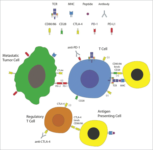
Figure 2. Immune checkpoints and checkpoint blockade. T lymphocytes, the effector cells of the immune system, recognize and are activated against metastatic tumor cells. This process is mediated via presentation of peptide antigens displayed on major histocompatibility complex (MHC) molecules of antigen presenting cells (APCs) to the T-cell receptors (TCRs), as well as binding of co-stimulatory molecules (such as CD28 on T cells binding to CD80 or CD86 on APCs). Immune checkpoints, such as CTLA-4 and PD-1, are induced on T cells following their activation. CTLA-4 is also expressed on regulatory T cells (Tregs). These checkpoints attenuate immune responses carried out by T cells. Tumor cells and Tregs utilize these molecules as a mechanism of immunosuppression within the tumor microenvironment. Immune checkpoint blockade is achieved using monoclonal antibodies directed against these molecules, thus relinquishing these mechanisms of immune inhibition. Evaluation of these therapies in patients with brain metastasis is currently underway.

Table 1. Selected clinical trials of immunotherapy for melanoma, lung, and breast cancer brain metastases.

Reprints and Corporate Permissions

Please note: Selecting permissions does not provide access to the full text of the article, please see our help page How do I view content?

To request a reprint or corporate permissions for this article, please click on the relevant link below:

Academic Permissions

Please note: Selecting permissions does not provide access to the full text of the article, please see our help page How do I view content?

Obtain permissions instantly via Rightslink by clicking on the button below:

If you are unable to obtain permissions via Rightslink, please complete and submit this Permissions form. For more information, please visit our Permissions help page.