Figures & data
Figure 1. Clustering of non-small cell lung cancers (NSCLC) according to the mRNA expression levels of CALR and that of metagenes indicating the presence of cytotoxic T cells (CTL) and dendritic cells (DC) and activated DC (aDC). (A) Expression z-scores of CALR, CTL and DC (combined in a pairwise fashion or combined altogether followed by clustering). (B) DC is replaced by aDC. Yellow dashed lines separate clusters and vertical color lines (black, red) define the clusters. BIC (Bayesian Information Criterion, defined as (−2) times the logarithm of the maximum likelihood, corrected by a factor that depends on the number of clusters and on the size of data) is indicated for each multidimensional clustering. (C) Three-dimensional representation of clusters obtained for the combined analysis of CALR, CTL and DC. The purple line represents the axis obtained by cluster dimension reduction (“MclustDR” function of R package Mclust). (D) Density plot representation of clusters, in the direction defined by purple axis in (C). (E) Three-dimensional representation of clusters obtained for the combined analysis of CALR, CTL and aDC. The purple line represents the axis obtained by cluster dimension reduction. (F) Density plot representation of clusters, in the direction defined by the purple axis in (F).
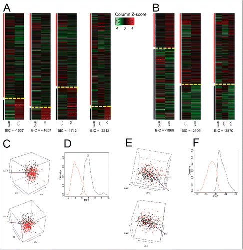
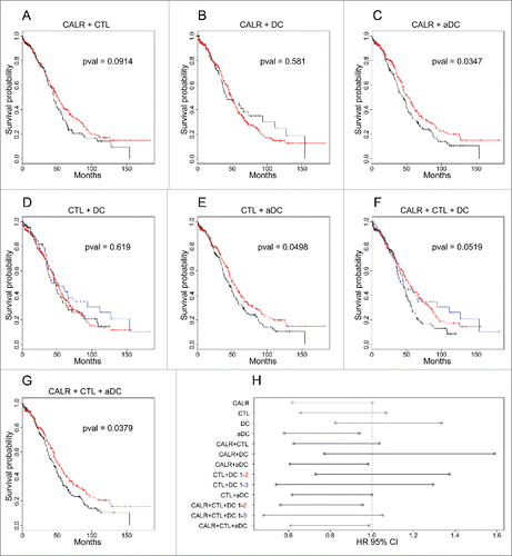
Figure 2. Survival curves and associated hazard ratios in NSCLC. The colors of the curves are related to the clusters indicated in . p values of survival curves were calculated by means of the log-rank test (A–G). Hazard ratios were obtained using the Cox model (H) for clusters defined by single (meta-)genes or their combination, as represented in Fig. S1.
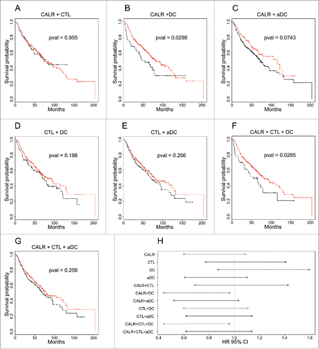
Figure 3. Clustering of ovarian cancers according to the mRNA expression levels of CALR and that of metagenes indicating the presence of cytotoxic T cells (CTL) and dendritic cells (DC) and activated DC (aDC). (A) Expression z-scores of CALR, CTL and DC (combined in a pairwise fashion or combined altogether followed by clustering). (B) DC is replaced by aDC. Yellow dashed lines separate clusters and vertical color lines (black, red, blue) define the clusters. BIC (Bayesian Information Criterion, defined as (−2) times the logarithm of the maximum likelihood, corrected by a factor that depends on the number of clusters and on the size of data) are indicated for each multidimensional clustering. (C) (respectively, E) Three-dimensional representation of clusters obtained for the combined analysis of CALR, CTL and DC (respectively, aDC). Purple lines represent the axes obtained by cluster dimension reduction (“MclustDR” function of R package mclust), the thick line for the first axis, the thin line for the second. (D) (respectively, F) scatter plot representation of clusters, in the directions defined by purple axes in (C) (respectively, F).
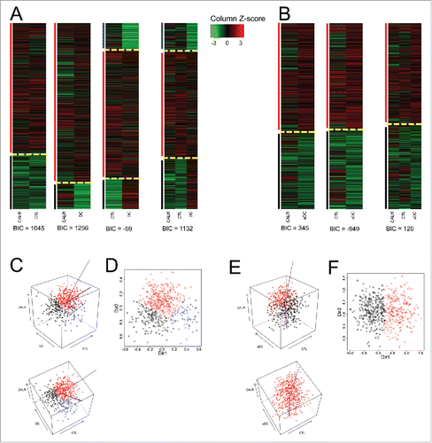
Figure 4. Survival curves and associated hazard ratios in ovarian cancer. The colors of curves are related to the clusters indicated in . p values of survival curves were calculated by means of the log-rank test (A–G). Hazard ratios were obtained using the Cox model (H) (including also cluster defined by single (meta-)gene, represented in Fig. S1). In the case of three clusters, the type of one-by-one comparison is indicated.