Figures & data
Figure 1. Ex vivo biodistribution of radiotracer Zr-89-DFO-PD-L1 mAb (10F.9G2) uptake in mice with MEER tumors treated by 2 Gy × 4 fractionated RT (A) or in mice with B16F10 tumors treated by 2 Gy × 4 fractionated RT (B) at 48 h after injection (3 d after completion of RT). Results are expressed in %ID/g and represent mean ± SEM (n = 5). *p < 0.05, paired t-test.
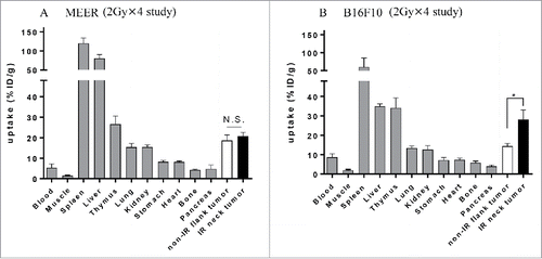
Figure 2. 2 Gy × 4 fractionated RT-induced PD-L1 upregulation in B16F10 was identified by PET/CT and its validity was corroborated by flow cytometry. (A) C57BL/6 mouse bearing MEER tumors or B16F10 tumors in two locations, neck and flank were treated with 2 Gy × 4 fractionated RT (only to the neck tumor) and anti PD-L1 PET/CT imaging and biodistribution study or flow cytometry were performed on day 20 after tumor implantation. (B) and (C) On day 1 after completion of RT, mice bearing MEER (B) or B16F10 (C) were injected Zr-89-DFO-PD-L1 mAb and PET/CT images were acquired at 48 h after injection. In-vivo or ex-vivo tracer uptake values (SUVmean and %ID/g, respectively) were compared between IR neck tumors and non-IR flank tumors (n = 5). Representative PET/CT images and tracer uptake values of IR and non-IR tumors were shown. (D) and (E) on day 3 after completion of RT, MEER tumors (D) or B16F10 tumors (E) were taken and flow cytometry analysis was performed. Representative flow cytometry histograms for PD-L1 expression and PD-L1 MFI of IR neck tumors and non-IR flank tumors of mice bearing MEER (n = 7, panel D) or B16F10 (n = 7), panel E). Sp, spleen; Li, liver. *p < 0.05, **p < 0.01; paired t-test.
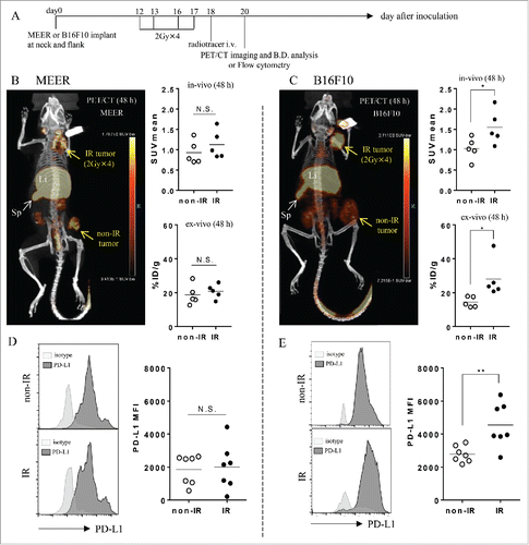
Table 1. Comparison of in vivo and ex vivo uptake values.
Figure 3. Ex vivo biodistribution of Zr-89-DFO-PD-L1 mAb uptake at 96 h after injection (at 4 d after completion of 2 Gy × 10 fractionated RT ± anti-PD-1 Ab). Biodistribution of radiotracer uptake in anti-PD-1 Ab treated mice and isotype control Ab treated mice with MEER tumors (n = 6 per group). Each mouse has two tumors in two locations, neck and flank, and fractionated RT was delivered only to the neck tumor. Results are expressed in %ID/g and represent mean ± SEM (n = 6). **p < 0.01; paired t-test.
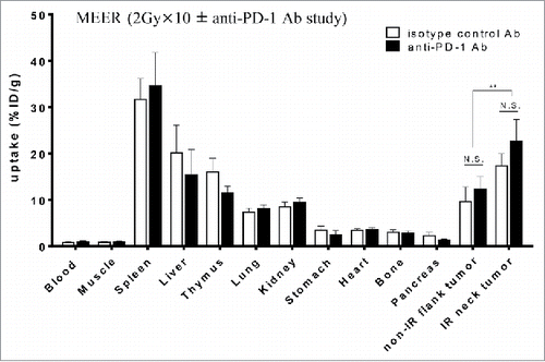
Figure 4. 2 Gy × 10 fractionated RT-induced PD-L1 upregulation in MEER was identified by PET/CT and its validity was corroborated by flow cytometry. (A) C57BL/6 mouse bearing MEER tumors in two locations, neck and flank were treated with fractionated RT (2 Gy × 10 only to the neck tumor) combined with anti-PD-1 Ab or isotype control Ab and PET/CT imaging and biodistribution study or flow cytometry were performed on day 33 after tumor implantation. (B) Tumor growth curves are presented as tumor volume measurements from day 10 to day 29 after tumor inoculation. Non-IR (isotype) tumors (n = 19), non-IR (anti-PD-1) tumors (n = 25), IR (isotype) tumors (n = 19) or IR (anti-PD-1) tumors (n = 25) were separately monitored. Data represent mean ± SEM (n = 5–25 per group). *p < 0.05, **p < 0.01; non-paired t-test. (C) RECIST outcome on day 29 after tumor inoculation (the last day of irradiation). (D) On the same day after completion of RT, mice bearing MEER treated with anti-PD-1 Ab or isotype control Ab were injected Zr-89-DFO-PD-L1 mAb and PET/CT images were acquired at 96 h after injection. In-vivo or ex-vivo tracer uptake values (SUVmean and %ID/g, respectively) were compared between non-IR (isotype) tumors and non-IR (anti-PD-1) tumors for analysis of anti-PD-1 Ab effect (n = 6), and thereafter compared between non-IR (isotype or anti-PD-1) tumors and IR (isotype or anti-PD-1) tumors for analysis of RT effect (n = 12). Representative PET/CT images and (E) SUVmean or %ID/g were shown. **p < 0.01; paired t-test. (F) On day 4 after completion of RT, MEER tumors were taken and flow cytometry analysis was performed. Representative flow cytometry histograms for PD-L1 expression and (G) the comparison of PD-L1 MFI between non-IR (isotype) tumors and non-IR (anti-PD-1) tumors for analysis of anti-PD-1 Ab effect (n = 5) and the comparison between non-IR (isotype or anti-PD-1) tumors and IR (isotype or anti-PD-1) tumors for analysis of RT effect (n = 10) were shown. Li, liver. *p < 0.05; paired t-test.
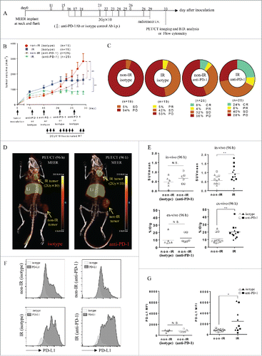
Figure 5. Flow cytometry analysis of CD45− cells or CD45+ cells in tumor tissues. Quantification of PD-L1 MFI and cell frequencies were measured. (A) Day 3 after completion of 2 Gy × 4 fractionated RT in MEER tumors. (B) Day 3 after completion of 2 Gy × 4 fractionated RT in B16F10 tumors. (C) Day 4 after completion of fractionated RT (2 Gy × 10) combined with anti-PD-1 Ab or its isotype control Ab in MEER tumors. Each mouse had two tumors in neck and flank, and only neck tumor was irradiated. *p < 0.05, **p < 0.01; paired t-test.
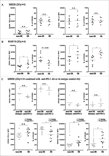
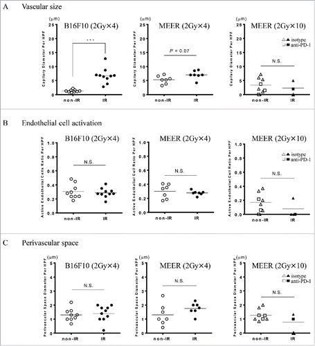
Figure 6. Quantitative immunohistochemical analysis of tumor vascular morphology. Morphologic parameters, including A, vascular size B, endothelial cell activation and C, perivascular space were measured to monitor the vascular changes that would influence tracer uptake. Marked vascular dilation after RT was seen in B16F10, however, no significant changes in the endothelial cell activation and the perivascular space were observed. ***p < 0.001; non-paired t-test.