Figures & data
Table 1. Clinicopathological and molecular features according to PDL1 expression.
Figure 1. PDL1, PDL2, PD1, CD3 and CD8 expression in gastric carcinoma by immunohistochemistry (× 200). PDL1 expression was evaluated based on staining in the cytoplasm and membrane of tumor cells and immune cells. A: Intense expression of PDL1 in tumor cells; B: Immune cells expressing PDL1 in the disseminated lymphocytes and macrophages; C: representative cytoplasmic expression of PDL2 in tumor tissues. D: PD1 expressed in the disseminated immune cells infiltrating the tumor tissues; E: The representative images of CD3+ T cells; F: The representative images of CD8+ T lymphocytes.
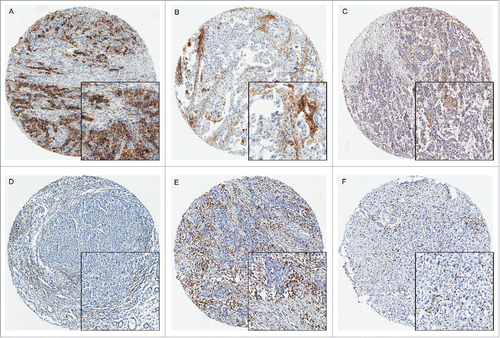
Table 2. Survival analysis of 1014 gastric cancers.
Figure 2. Kaplan-Meire curves stratified by PDL1, CD3, and CD8 expression. PDL1 expression in tumor cells predicted better survival in GC patients using cut-off value of IRS for 2 and 5 as 3 groups (A, PDL1TU: PDL1 expression in tumor cells) or in the immune cells (B, PDL1IM: PDL1 expression in immune cells). CD3+ (C) or CD8+ (D) T cells both associated with better outcome in a density dependent manner.
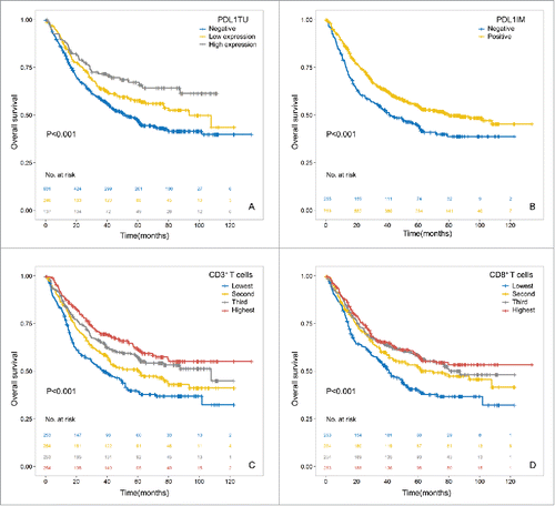
Figure 3. Prognostic significance of PDL1 expression (A) in tumor cells as well as the intersection of PDL1 expression and CD3+ T cell infiltration (B) in subgroups of different pTNM stages. Using the median of CD3 density as the cutoff value and PDL1 membrane expression in tumor cells, the patients were divided into 2 groups, one with CD3 density lower than median and PDL1 negative expression, and the other group consisted with the rest patients.
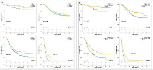
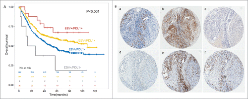
Figure 4. PDL1 expression associated with better outcome in the subgroup of EBV+ and EBV- GC patients (A). PDL1, PDL2, PD1, CD3 and CD8 expression in the identical EBV infected gastric cancer tissue (× 200, B). In situ hybridization of EBER (a) was shown in the identical patients together with PDL1 (b), PDL2 (c) and PD1 (d) expression in GC tumor tissues and CD3 (e) and CD8 (f) in intratumoral lymphocytes.