2,028
Views
37
CrossRef citations to date
0
Altmetric
Original Research

IL-17 induces antitumor immunity by promoting beneficial neutrophil recruitment and activation in esophageal squamous cell carcinoma

, , , , , , , , , , , & show all
Article: e1373234 | Received 22 Jun 2017, Accepted 25 Aug 2017, Published online: 21 Sep 2017

Figures & data

Figure 1. Infiltration of MPO+ neutrophils into tumor nests and its association with survival in ESCC patients. The MPO+ neutrophils in ESCC surgical specimens were detected by immunohistochemistry. According to the median count of MPO+ cells, the patients were divided into a high MPO+ infiltration group (A) and a low MPO+ infiltration group (B). Upper panels, × 200 magnification; lower panels, × 400 magnification. (C) Survival analysis shows that overall survival (OS) was significantly improved in the high MPO+ infiltration group compared to the low MPO+ infiltration group.
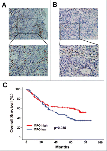
Figure 1. Infiltration of MPO+ neutrophils into tumor nests and its association with survival in ESCC patients. The MPO+ neutrophils in ESCC surgical specimens were detected by immunohistochemistry. According to the median count of MPO+ cells, the patients were divided into a high MPO+ infiltration group (A) and a low MPO+ infiltration group (B). Upper panels, × 200 magnification; lower panels, × 400 magnification. (C) Survival analysis shows that overall survival (OS) was significantly improved in the high MPO+ infiltration group compared to the low MPO+ infiltration group.

Table 1. Association of MPO+ neutrophile count and clinical characteristics in ESCC.

Figure 2. Correlation between MPO+ neutrophil infiltration and IL-17-producing cells in intratumoral areas of ESCC. (A) Immunohistochemical staining of MPO+ cells and IL-17+ cells in the same tumor tissues (Upper panel: sample 1; lower panel: sample 2). Original magnification, × 200. (B) Linear regression analyses suggest that there is a significantly positive correlation between the densities of MPO+ cells and IL-17+ cells in ESCC tissues. (C) Increased infiltration of both MPO+ cells and IL-17+ cells indicated better overall survival in ESCC patients.
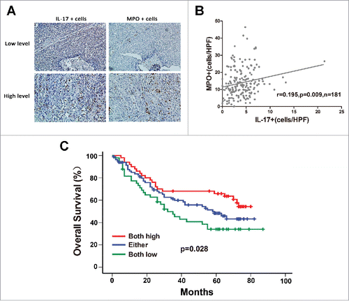
Figure 2. Correlation between MPO+ neutrophil infiltration and IL-17-producing cells in intratumoral areas of ESCC. (A) Immunohistochemical staining of MPO+ cells and IL-17+ cells in the same tumor tissues (Upper panel: sample 1; lower panel: sample 2). Original magnification, × 200. (B) Linear regression analyses suggest that there is a significantly positive correlation between the densities of MPO+ cells and IL-17+ cells in ESCC tissues. (C) Increased infiltration of both MPO+ cells and IL-17+ cells indicated better overall survival in ESCC patients.

Figure 3. IL-17 recruits neutrophils by stimulating tumor cell-derived CXC chemokine production in ESCC. (A) The supernatants of IL-17-treated EC109 cells (IL17_ EC109) significantly increased the migration of neutrophils compared with IL-17 plus EC109 cell supernatant (IL17+ EC109), EC109 cell supernatant, IL-17, or medium alone. Data are presented from five separate experiments using one donor's neutrophils. (B) Fold changes in CXC chemokine mRNA levels in IL-17-treated EC109 cells compared to untreated EC109 cells were quantified by qRT-PCR. Data from three separate experiments are presented. (C) ELISA analysis showed that IL-17 stimulated EC109 cells to secrete more chemokines CXCL2 and CXCL3. Data from three separate experiments are presented. (D) Silencing of CXCL2 and CXCL3, but not of CXCL4 or CXCL16 in IL-17-treated EC109 cells attenuated the migration of neutrophils (siControl vs.siCXCL2, siCXCL3, siCXCL2+siCXCL3, siCXCL4, or siCXCL16). Data are presented from three separate experiments using one donor's neutrophils. (E and F) Immunohistochemical analysis of the association between CXCL2/ CXCL3 expression and the number of IL-17+ cells or MPO+ cells in 181 ESCC tumor samples. Representative examples of IL-17, MPO and CXCL2 or CXCL3 staining in the same tumor tissues are shown in (E). (F) Statistical analysis shows a significantly positive correlation between IL-17 and CXCL2 /CXCL3 expression, and between CXCL2 /CXCL3 expression and the number of MPO+ cells. (G) Overall survival was significantly enhanced in IL-17high MPOhigh CXCL2high or IL-17high MPOhigh CXCL3high patients compared with IL-17low MPOlow CXCL2low or IL-17low MPOlow CXCL3low patients. * P < 0.05, ** P < 0.01, *** P < 0.001.
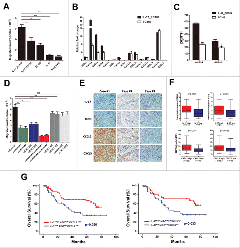
Figure 3. IL-17 recruits neutrophils by stimulating tumor cell-derived CXC chemokine production in ESCC. (A) The supernatants of IL-17-treated EC109 cells (IL17_ EC109) significantly increased the migration of neutrophils compared with IL-17 plus EC109 cell supernatant (IL17+ EC109), EC109 cell supernatant, IL-17, or medium alone. Data are presented from five separate experiments using one donor's neutrophils. (B) Fold changes in CXC chemokine mRNA levels in IL-17-treated EC109 cells compared to untreated EC109 cells were quantified by qRT-PCR. Data from three separate experiments are presented. (C) ELISA analysis showed that IL-17 stimulated EC109 cells to secrete more chemokines CXCL2 and CXCL3. Data from three separate experiments are presented. (D) Silencing of CXCL2 and CXCL3, but not of CXCL4 or CXCL16 in IL-17-treated EC109 cells attenuated the migration of neutrophils (siControl vs.siCXCL2, siCXCL3, siCXCL2+siCXCL3, siCXCL4, or siCXCL16). Data are presented from three separate experiments using one donor's neutrophils. (E and F) Immunohistochemical analysis of the association between CXCL2/ CXCL3 expression and the number of IL-17+ cells or MPO+ cells in 181 ESCC tumor samples. Representative examples of IL-17, MPO and CXCL2 or CXCL3 staining in the same tumor tissues are shown in (E). (F) Statistical analysis shows a significantly positive correlation between IL-17 and CXCL2 /CXCL3 expression, and between CXCL2 /CXCL3 expression and the number of MPO+ cells. (G) Overall survival was significantly enhanced in IL-17high MPOhigh CXCL2high or IL-17high MPOhigh CXCL3high patients compared with IL-17low MPOlow CXCL2low or IL-17low MPOlow CXCL3low patients. * P < 0.05, ** P < 0.01, *** P < 0.001.

Figure 4. IL-17 intensifies the antitumor activity of neutrophils against ESCC tumor cells by enhancing the expression of cytotoxic molecules. (A) LDH assays show that IL-17 promotes the cytotoxicity of neutrophils against EC109 cells. Data are presented from three separate experiments using one donor's neutrophils. (B) IL-17 increases ROS production in neutrophils determined by flow cytometric analysis. The flow cytometry histogram plot for depicting ROS generation is shown on the left. The corresponding mean fluorescence intensity (MFI) is presented on the right from three separate experiments using one donor's neutrophils. (C) ELISA analysis shows that IL-17 significantly enhances the release of TRAIL and IFN-γ but does not affect NE or TNF-α production from neutrophils. Data are presented from four separate experiments using one donor's neutrophils. (D-G) Flow cytometric analysis was conducted to evaluate the effect of IL-17 on the expression of MPO, CXCR1, and CXCR2 on neutrophils. Data from four separate healthy donors' neutrophils are presented. (D) The gating routine for CD16+ neutrophils from healthy donors. (E-G) The dot plots represent the MPO+ cell subset (E), the CXCR1+ cell subset (F) and the CXCR2+ cell subset (G) gating on the CD16+fraction, and the statistical analysis suggests that IL-17 can up-regulate MPO expression in neutrophils but does not affect CXCR1 or CXCR2 expression. * P < 0.05, ** P < 0.01, *** P < 0.001.
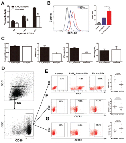
Figure 4. IL-17 intensifies the antitumor activity of neutrophils against ESCC tumor cells by enhancing the expression of cytotoxic molecules. (A) LDH assays show that IL-17 promotes the cytotoxicity of neutrophils against EC109 cells. Data are presented from three separate experiments using one donor's neutrophils. (B) IL-17 increases ROS production in neutrophils determined by flow cytometric analysis. The flow cytometry histogram plot for depicting ROS generation is shown on the left. The corresponding mean fluorescence intensity (MFI) is presented on the right from three separate experiments using one donor's neutrophils. (C) ELISA analysis shows that IL-17 significantly enhances the release of TRAIL and IFN-γ but does not affect NE or TNF-α production from neutrophils. Data are presented from four separate experiments using one donor's neutrophils. (D-G) Flow cytometric analysis was conducted to evaluate the effect of IL-17 on the expression of MPO, CXCR1, and CXCR2 on neutrophils. Data from four separate healthy donors' neutrophils are presented. (D) The gating routine for CD16+ neutrophils from healthy donors. (E-G) The dot plots represent the MPO+ cell subset (E), the CXCR1+ cell subset (F) and the CXCR2+ cell subset (G) gating on the CD16+fraction, and the statistical analysis suggests that IL-17 can up-regulate MPO expression in neutrophils but does not affect CXCR1 or CXCR2 expression. * P < 0.05, ** P < 0.01, *** P < 0.001.

Figure 5. IL-17 is involved in neutrophil recruitment and activation against ESCC tumor growth in vivo. (A) MTS assays indicated that overexpression of IL-17 does not significantly affect ex vivo EC109 cell proliferation. Data from three separate experiments are presented. (B) IL-17 secretion from EC109/IL-17 cells or EC109/Vector cells was detected by ELISA. Data from three separate experiments are presented. (C and D) Photographs of the EC109 cell xenograft model (C) and dissected tumors from nude mice (D) (n = 6). (E) The tumor growth curves for each group are presented. IL-17 overexpression markedly reduced the ESCC tumor growth rate when the nude mice were treated with human neutrophils via tail vein injection. (F) The weights of tumors from each group are shown. The final tumor weights were much lower in the group with overexpression of IL-17 plus human neutrophil infusions. (G and H) Immunohistochemical analysis for quantification of IL-17 expression, CXCL2 and CXCL3 expression, MPO+ cell infiltration, and the proliferation marker Ki-67 expression in the dissected tumors from nude mice. (G) Representative pictures of IL-17, CXCL2, CXCL3, MPO and Ki-67 staining at tumor sites for each group are shown. Original magnification, × 200. (H) The statistical analysis shows that CXCL2 and CXCL3 expression is much higher in the IL-17 overexpressing group. The tumors from the IL-17 overexpression plus human neutrophil infusion group have higher MPO+ cell infiltration and lower Ki-67 expression. Data from six separate experiments are presented. ** P < 0.01, *** P < 0.001; ns, no significance.
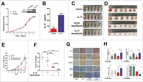
Figure 5. IL-17 is involved in neutrophil recruitment and activation against ESCC tumor growth in vivo. (A) MTS assays indicated that overexpression of IL-17 does not significantly affect ex vivo EC109 cell proliferation. Data from three separate experiments are presented. (B) IL-17 secretion from EC109/IL-17 cells or EC109/Vector cells was detected by ELISA. Data from three separate experiments are presented. (C and D) Photographs of the EC109 cell xenograft model (C) and dissected tumors from nude mice (D) (n = 6). (E) The tumor growth curves for each group are presented. IL-17 overexpression markedly reduced the ESCC tumor growth rate when the nude mice were treated with human neutrophils via tail vein injection. (F) The weights of tumors from each group are shown. The final tumor weights were much lower in the group with overexpression of IL-17 plus human neutrophil infusions. (G and H) Immunohistochemical analysis for quantification of IL-17 expression, CXCL2 and CXCL3 expression, MPO+ cell infiltration, and the proliferation marker Ki-67 expression in the dissected tumors from nude mice. (G) Representative pictures of IL-17, CXCL2, CXCL3, MPO and Ki-67 staining at tumor sites for each group are shown. Original magnification, × 200. (H) The statistical analysis shows that CXCL2 and CXCL3 expression is much higher in the IL-17 overexpressing group. The tumors from the IL-17 overexpression plus human neutrophil infusion group have higher MPO+ cell infiltration and lower Ki-67 expression. Data from six separate experiments are presented. ** P < 0.01, *** P < 0.001; ns, no significance.
Supplemental material

Reprints and Corporate Permissions

Please note: Selecting permissions does not provide access to the full text of the article, please see our help page How do I view content?

To request a reprint or corporate permissions for this article, please click on the relevant link below:

Academic Permissions

Please note: Selecting permissions does not provide access to the full text of the article, please see our help page How do I view content?

Obtain permissions instantly via Rightslink by clicking on the button below:

If you are unable to obtain permissions via Rightslink, please complete and submit this Permissions form. For more information, please visit our Permissions help page.