
Figures & data
Figure 1. EV characterization by western blot and nanoparticle tracking analysis. (A) EV marker expression analysis for HSP70, CD63, TSG101, Syntenin, CD9, CD81, Cytochrome C (Cyt C) in EV fractions from two healthy persons (NP1, NP2), three BC patients pre-NACT (1-3) and post-NACT (4-6). In all analyses, HEK cell culture supernatant derived EVs served as positive control, for Cyt C detection MCF-7 cell lysate (*) was used as control. (B) The EV size [median (interquartile range) nm] did not differ pre- and post-NACT (Mann-Whitney test). (C) Healthy females presented lower EV levels [median (interquartile range) 109/ml] compared to BC patients pre- and post-NACT. EV levels increased post-NACT in BC patients (Mann-Whitney test).
![Figure 1. EV characterization by western blot and nanoparticle tracking analysis. (A) EV marker expression analysis for HSP70, CD63, TSG101, Syntenin, CD9, CD81, Cytochrome C (Cyt C) in EV fractions from two healthy persons (NP1, NP2), three BC patients pre-NACT (1-3) and post-NACT (4-6). In all analyses, HEK cell culture supernatant derived EVs served as positive control, for Cyt C detection MCF-7 cell lysate (*) was used as control. (B) The EV size [median (interquartile range) nm] did not differ pre- and post-NACT (Mann-Whitney test). (C) Healthy females presented lower EV levels [median (interquartile range) 109/ml] compared to BC patients pre- and post-NACT. EV levels increased post-NACT in BC patients (Mann-Whitney test).](/cms/asset/21d20d68-e16a-4209-9d8f-d5898636b1a4/koni_a_1376153_f0001_b.gif)
Table 1. Association of pre- and post-NACT EV levels with clinical parameters.
Table 2. Post-NACT EV level cut-off discriminates between CTC subtypes. BC patients having post-NACT EV levels below the cut-off had a higher incidence of SL-CTCs, ALDH-1 positivity, ERCC1 positivity and ALDH-1/ERCC1 positivity. Data was evaluated by Fisher's exact test.
Figure 2. ROC analyses for cut-off determination for EV levels pre- and post-NACT and Kaplan-Meier survival analysis regarding 3y-PFS and OS. (A)/(C) By ROC analysis a consistent cut-off value of 2484 ×109/ml for EV levels post-NACT was obtained for 3y-PFS and OS. (B/D) In Kaplan Meier analysis combined with the log rank test, BC patients having EV levels post-NACT above the determined cut-off value had a significantly reduced 3y-PFS and OS.
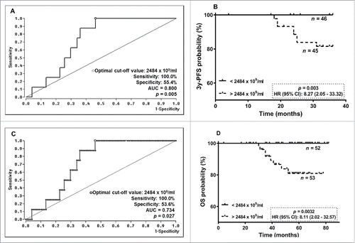
Figure 3. Association between CTC subtypes and EV levels post-NACT. EV concentrations post-NACT were reduced if the following CTC subpopulations were detectable: (A) SL-CTCs, (B) ALDH-1 positive, (C) ERCC1 positive and (D) ALDH-1/ERCC1 positive. Data is shown as EV levels [median (interquartile range) 109/ml] and was evaluated by Mann-Whitney test. The dotted line indicates the cut-off value of 2484 ×109 EVs/ml.
![Figure 3. Association between CTC subtypes and EV levels post-NACT. EV concentrations post-NACT were reduced if the following CTC subpopulations were detectable: (A) SL-CTCs, (B) ALDH-1 positive, (C) ERCC1 positive and (D) ALDH-1/ERCC1 positive. Data is shown as EV levels [median (interquartile range) 109/ml] and was evaluated by Mann-Whitney test. The dotted line indicates the cut-off value of 2484 ×109 EVs/ml.](/cms/asset/7daa29da-1451-47b3-87d1-6b5e1ee17ec1/koni_a_1376153_f0003_b.gif)
Table 3. Patient characteristics.