
Figures & data
Figure 1. mIL-21 combined with either mCTLA-4 or mPD-1 mAb in the EMT-6 mammary carcinoma tumor model. (A) Antitumor activity of mIL-21 (50 μg/mouse) and mCTLA-4 mAb (clone UC10-4F10; 400 μg/mouse) and (B) antitumor activity of mIL-21 (50 μg/mouse) and PD-1 mAb (clone 4H2-mIgG1; 200 μg/mouse), when administered alone or in combination on the days indicated in the table. Median tumor volumes (left hand panels) and individual tumor volumes (right hand panels) are plotted vs. days post implant for control (untreated; circles), mCTLA-4 or mPD-1 mAb- (squares), mIL-21- (triangles), or mIL-21 + mAb (inverted triangles)-treated groups. CR = complete regression. Asterisks (*, **) indicate p < 0.05 or p < 0.01, respectively, for differences between the mCTLA-4 mAb + mIL-21 combination group and the mCTLA-4 mAb group (p < 0.05), or the combination group and either the control or mIL-21 group (p < 0.01) for ‘treatment effect’ by 2-way repeated-measures ANOVA. Data are representative of results from two separate studies.
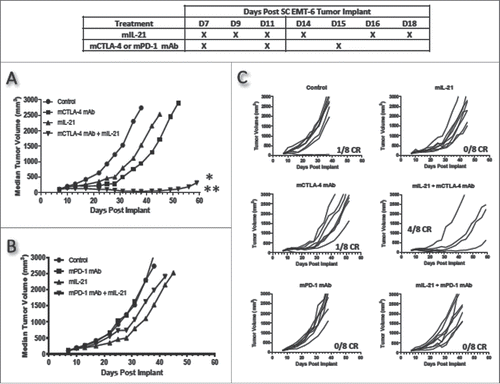
Figure 2. mIL-21 combined with mCTLA-4 or mPD-1 mAb in the IV B16-F10 lung metastatic melanoma model. (A) Antitumor activity of mIL-21 (75 μg/mouse) and mCTLA-4 mAb (9D9-mIgG2b; 300 μg/mouse), alone or in combination, on day 20 post-tumor cell implant. (B) Antitumor activity of mIL-21 (75 μg/mouse) and mPD-1 mAb (4H2-mIgG1; 300 μg/mouse), alone or in combination, on day 20 post-cell implant. Mean values +/− SEM are shown. Asterisks (*, **, ***) indicate p < 0.05, 0.01 or 0.001, respectively, for differences between groups by one way ANOVA. There were no other statistically significant differences between groups. Data are representative of results from two separate studies.
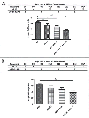
Figure 3. mIL-21 combined with either mCTLA-4 or mPD-1 mAb in the MC38 colon carcinoma tumor model. (A) Antitumor activity of mIL-21 (200 μg/mouse) and mCTLA-4 mAb (9D9-mIgG2b; 200 μg/mouse) administered alone or in combination on the days indicated in the table. (B) Antitumor activity of mIL-21 (50 μg/mouse) and PD-1 mAb (4H2-mIgG1; 200 μg/mouse), administered alone or in combination on the days indicated. Median tumor volumes (left hand panels) and individual tumor volumes (right hand panels) are plotted vs. days post-tumor cell implant for mIgG1 control- (crosses), mIgG1 + mIL-21- (triangles), mCTLA-4 or mPD-1 mAb- (squares), or mIL-21 + mAb- (diamonds)-treated groups. For Panel A, asterisks (**, ***) indicate p < 0.01 or p < 0.001, respectively, for differences between the mCTLA-4 mAb + mIL-21 combination group and either the mCTLA-4 mAb group or mIL-21 group (each comparison is p < 0.01), or the combination group and the mIgG control group (p < 0.001) for ‘treatment effect’ by 2-way repeated measures ANOVA. For Panel B, asterisks (*, ***) indicate p < 0.05 or p < 0.001, respectively, for differences between the mPD-1 mAb + mIL-21 combination group and either the mPD-1 mAb group or mIL-21 group (each comparison is p < 0.05), or the combination group and the control group (p < 0.001) for ‘treatment effect’ by 2-way repeated measures ANOVA. CR = complete regression. Data are representative of results from two separate studies.
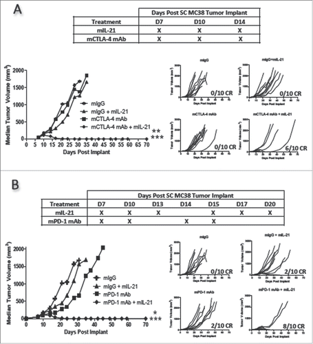
Figure 4. Combination treatment enhances CD8+ T cell infiltration into tumors in the MC38 colon carcinoma model. (A) TILs were isolated from MC38 tumors on day 13 after implantation and analyzed by flow cytometry. The percentage of CD8+ cells in the total CD45+ population and the percentage of Ki67+CD8+ T cells for each treatment group are plotted. Each symbol represents data from one mouse in the group and mean values are indicated with horizontal lines. Asterisks (*) indicate p < 0.05 for differences between the groups indicated by 1-way ANOVA. (B-G) Tumors isolated in A were frozen in OCT, sectioned, and stained for CD8-expressing cells. Treatment groups were as follows: (B) PBS-treated control, (C) 50 µg mIL-21, 3 times per week, 6 total doses, (D) 200 µg mCTLA-4 mAb (9D9-mIgG2b), every 4 days, 2 total doses, (E) 50 µg mIL-21 plus 200 µg mCTLA-4 mAb, every 4 days, 2 total doses. (F) 200 µg mPD-1 mAb (4H2-mIgG1; 3 doses). (G) 50 µg mIL-21 (6 doses) plus 3 doses of 200 µg mPD-1 mAb. Bar = 100 µm. Study was conducted once.
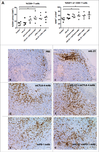
Figure 5. Memory T cell subsets in TILs from MC38 tumors in mice treated with mIL-21 +/− mPD-1 or mCTLA-4 mAbs. TILs from mice implanted SC with MC38 tumor cells and treated as described in with PBS, mIL-21, mCTLA-4 mAb (9D9-mIgG2b), mPD-1 mAb (4H2-mIgG1), or mIL-21 + mPD-1 or mCTLA-4 mAbs were isolated on study day 13, stained with various markers of immune cell subsets, and evaluated by flow cytometry. The %CD44-CD62 L+ (upper left), %CD44+CD62 L+ (TCM; upper right), %CD44+CD62 L- (TEM; lower left), and %CD44-CD62 L- (lower right) among live CD45+ (A) CD4+ or (B) CD8+ cells, are shown for each treatment group, indicated on the x-axes. Each symbol represents data from one mouse in the group and mean values are indicated with horizontal lines. Asterisks (*, **) indicate p < 0.05 or p < 0.01, respectively, for differences between the groups indicated by 1-way ANOVA. Study was conducted once.
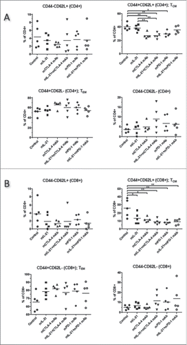
Figure 6. Addition of mIL-21 improves antitumor activity of mCTLA-4 mAb in the CT26 colon carcinoma model. Antitumor activity of mIL-21 (50 μg/mouse) and anti-mCTLA-4 mAb (clone UC10-4F10; 400 μg/mouse) when administered alone or in combination on the days indicated in the table. Median tumor volumes (left hand panel) and individual tumor volumes (right hand panels) are shown. CR = complete regression. Asterisks (*, **) indicate p < 0.05 or p < 0.01, respectively, for differences between the mCTLA-4 mAb group and either the control group or mIL-21 group (each comparison is p < 0.05), or the mCTLA-4 + mIL-21 combination group and either the control group or mIL-21 group (each comparison is p < 0.01) for ‘treatment effect’ by 2-way repeated measures ANOVA. Data are representative of results from two separate studies.
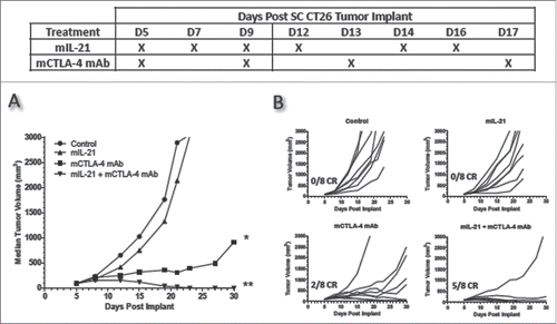
Figure 7. TIL immunophenotypes in CT26 tumors from mice treated with mIL-21 +/− mPD-1 or mCTLA-4 mAbs. TILs from mice implanted SC with CT26 tumor cells and treated with control mIgG, mIL-21, mCTLA-4 mAb (9D9-mIgG2b), mPD-1 mAb (4H2-mIgG1), or mIL-21 + mPD-1 or mCTLA-4 mAbs were isolated on study day 16, stained with various markers of immune cell subsets, and evaluated by flow cytometry. The (A) %CD8+ of live CD45+, (B) %CD25+FoxP3+ of CD4+, (C) %CD335+ of live CD45+, and (D) %PD-1+CD69- of CD8+ cells, are shown for each treatment group, indicated on the x-axes. The (E) ratios of the % CD8+ T cells of live CD45+ (Teff) to the % CD4+CD25+ of live CD45+ (Tregs) are plotted. Each symbol represents data from one mouse in the group and mean values are indicated with horizontal lines. Asterisks (*, **, ***, ****) indicate p < 0.05, p < 0.01, p < 0.001 or p < 0.0001, respectively, for differences between the groups indicated by 1-way ANOVA. Study was conducted one time.
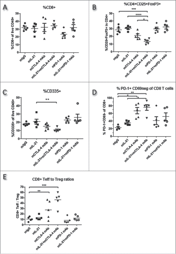
Figure 8. Effect of immune cell subset depletion on mIL-21 + CTLA-4 or PD-1 blockade in the MC38 and CT26 models. (A) Mice implanted SC with (A) MC38 or (B) CT26 tumor cells were injected IP with PBS (upper left), or depleting mAbs directed against CD4+ (upper right), CD8+ (lower left), or NK (lower right) cells on days 3, 9, and 16 post-tumor implant. Two days after initiating depleting mAb treatment, the mice were administered PBS (x's), mCTLA-4 mAb (9D9-mIgG2b, 200 μg/mouse; filled circles), mIL-21 (50 μg/mouse; filled diamonds), a combination of mIL-21 and mCTLA-4 mAb (open circles), mPD-1 mAb (4H2-mIgG1, 200 μg/mouse; filled squares), or a combination of mIL-21 and mPD-1 mAb (open squares). Median tumor volumes (mm3) are plotted vs. days post tumor cell implant. Data are representative of results from two separate studies. See Supplemental Table 1 for statistical analyses.
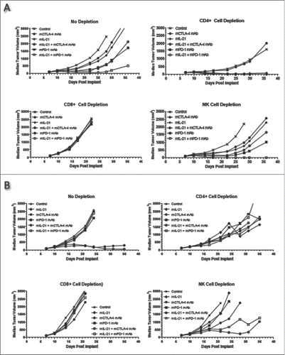
Table 1. Summary of antitumor efficacy of mIL-21+mCTLA-4 mAb or mPD-1 mAb.
Table 2. Experimental Design.