3,697
Views
59
CrossRef citations to date
0
Altmetric
Original Research

Prognostic relevance of tumor-infiltrating lymphocytes and immune checkpoints in pediatric medulloblastoma

, , , , ORCID Icon, , , , , & show all
Article: e1398877 | Received 08 Sep 2017, Accepted 24 Oct 2017, Published online: 27 Nov 2017

Figures & data

Table 1. Patient characteristics.

Figure 1. Distribution of tumor infiltrating lymphocytes in pediatric medulloblastoma. Distribution of CD3+ T-cells in pediatric medulloblastoma resembles two distinct patterns i.e. perivascular (left panel) and intratumoral (right panel) that often coincide. Scale bar equals 100 μm.
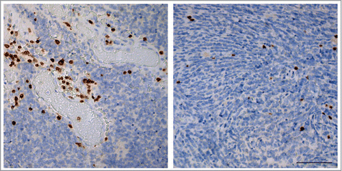
Figure 1. Distribution of tumor infiltrating lymphocytes in pediatric medulloblastoma. Distribution of CD3+ T-cells in pediatric medulloblastoma resembles two distinct patterns i.e. perivascular (left panel) and intratumoral (right panel) that often coincide. Scale bar equals 100 μm.

Figure 2. Expression of immune checkpoints and evasion markers in pediatric medulloblastoma. A) Immunohistochemical staining of immune (evasion) markers HLA-A, HLA-B and CD1 d in one case of pediatric medulloblastoma demonstrating that expression of all these markers is absent compared to endothelium and TILs. B) Examples of SerpinB1, SerpinB4 expression in pediatric medulloblastoma. SerpinB9 expression was not detected in pediatric medulloblastoma. C) Expression PD-L1 was not detected in pediatric medulloblastoma by immunohistochemistry. PD-1 positive TILs (arrowheads) are predominantly present at the peripheral zone of the tumor. Positive controls: placenta (PD-L1) and tonsil (PD-1). Scale bar equals 100 μm.
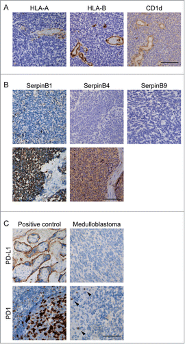
Figure 2. Expression of immune checkpoints and evasion markers in pediatric medulloblastoma. A) Immunohistochemical staining of immune (evasion) markers HLA-A, HLA-B and CD1 d in one case of pediatric medulloblastoma demonstrating that expression of all these markers is absent compared to endothelium and TILs. B) Examples of SerpinB1, SerpinB4 expression in pediatric medulloblastoma. SerpinB9 expression was not detected in pediatric medulloblastoma. C) Expression PD-L1 was not detected in pediatric medulloblastoma by immunohistochemistry. PD-1 positive TILs (arrowheads) are predominantly present at the peripheral zone of the tumor. Positive controls: placenta (PD-L1) and tonsil (PD-1). Scale bar equals 100 μm.

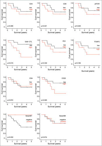
Figure 3. TILs and survival of pediatric medulloblastoma patients. Survival analysis of pediatric medulloblastoma patients stratified for amount of TILs. High and low number of TILs was determined by median split.

Figure 3. TILs and survival of pediatric medulloblastoma patients. Survival analysis of pediatric medulloblastoma patients stratified for amount of TILs. High and low number of TILs was determined by median split.