
Figures & data
Table 1. Patient profiles and treatment response.
Figure 1. Genomic alterations identified* Top Panel: Total alterations (N = 62) identified by NGS on initial biopsy (N = 8 biopsies). Bottom Panel: Total alterations (N = 77) identified by NGS on initial and subsequent biopsies (N = 10 biopsies). *Some patients had multiple alterations in the same gene (i.e. TP53 and PTCH1).
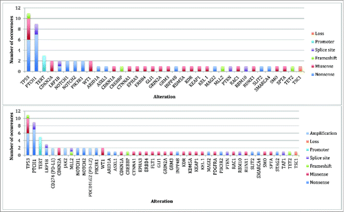
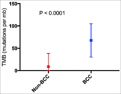
Figure 2. Mean tumor mutational burden for cancers other than basal cell carcinoma (N = 1,637) vs. basal cell carcinoma (N = 9 biopsies with available data). P value calculated using Mann Whitney U test. Squares represent mean TMB. Bars represent the standard deviation of the mean. Abbreviations: BCC = basal cell carcinoma; mb = megabase; TMB = tumor mutational burden.
Supplemental material