Figures & data
Figure 1. Sequential analysis of peripheral blood T cells by high-resolution flow cytometry and TCRα, V, and J assignment (A) Results from an acute type ATL patient who received both conventional chemotherapy mLSG15 (A-1 to A-2) and moga (A-2 to A-3). Flow cytometric scattergrams illustrating percentages of CD4+/8+ and CADM1+CD7− (ATL) cells. For TCR repertoire analysis, 3D graphs show the frequencies of TRAV/J clones. ATL cells with a CADM1+CD7− phenotype decreased remarkably and reconstitution of polyclonal T cells was observed after moga treatment (A-3); however, 7 months later, a CADM1+CD7− cell population and the TRAV21/J49 clone reappeared and polyclonal T cells were suppressed (A-4). (B) Results from another acute type ATL patient who received mLSG15 (B-1 to B-2) and moga (B-2 to B-3). After moga treatment, PBMCs were sorted into CD4+ and CD8+ T cells, and subjected to TCR repertoire analysis (B-3 (CD4) and B-3 (CD8)). Data are representative of five independent patients. N.D.: not detected.
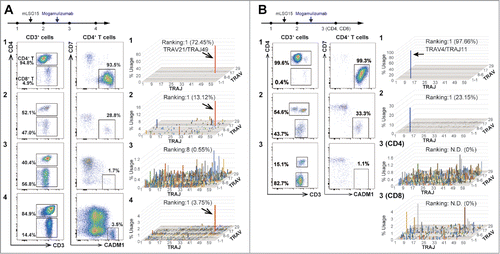
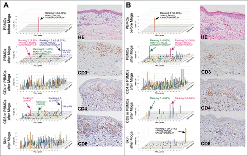
Figure 2. Peripheral blood T cells and cutaneous infiltrating T cells do not share the same major T cell clones after treatment with moga (A, B) PBMCs, sorted CD4+/CD8+ T cells, and skin tissues were subjected to TCR repertoire analysis in two ATL cases with moga-related skin rash. 3D graphs show the frequencies of TRAV/J clones. In panel A, the blue bar consists of three clones with different amino acid sequences in the CDR3 region, but sharing TRAV24/J52. H&E and CD3/4/8 staining of the samples of tissue from the skin rash are also shown. Data are representative of four independent patients.