
Figures & data
Figure 1. Cancer cells under T cell immune attack upregulate cell surface CD47
(a) Representative flow cytometric contour plots of CD47 expression (MFI) in Annexin-Vneg PC-3 target cells after co-culturing with HLA-matched CMVpp65-specific T cells at indicated E:T cell ratios in the presence (or absence) of Retarg-1 for 48 h. (b) Induction of cancer cell death in PC-3 target cells (% Annexin-Vpos cells) by CMVpp65-specific T cells. Relative fold increase of (c) CD47 and (d) PD-L1 cell surface expression on Annexin-Vneg PC-3 target cells at indicated E:T cell ratios. (e) CD47 cell surface presence in Annexin-Vneg/EpCAMpos target cells (PC-3, NCI-H292, OvCAR-3 and FaDu) after treatment with CMVpp65-specific T cells in the presence (or absence) of Retarg-1 at an E:T cell ratio of 1:1. (f) CD47 cell surface presence in Annexin-Vneg/EpCAMpos target cells after treatment with PBMCs in the presence (or absence) of T cell-redirecting (anti-EpCAM/anti-CD3) bsAb BIS1 at an E:T cell ratio of 2:1. Graphs represent mean ± SD. All experiments were analyzed by flow cytometry. Statistical analysis in graphs E and F was performed using unpaired T-test (ns = not significant, *p < .05, **p < .01, ***p < .001).
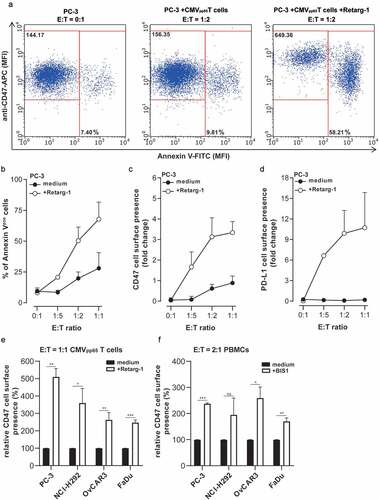
Figure 2. Upregulation of CD47 expression enhances resistance of cancer cells to T cell-mediated cytotoxicity
(a) Induction of cancer cell death (% Annexin-Vpos/PIpos) in Retarg-1-decorated PC-3 or (b) PC-3.CD47KO cells when treated with HLA-matched CMVpp65-specific T cells at indicated E:T cell ratios. (c) The percentage of Annexin-V positivity in PC-3 target cells ectopically overexpressing CD47 (PC-3.CD47high, open circles and open boxes) versus empty vector-transduced PC-3 cells (PC-3.ev, solid black circles and solid black boxes) after treatment with CMVpp65-specific T cells in the presence of Retarg-1. (d) IFNγ and (e) granzyme B levels in culture supernatants of (c) were measured by ELISA. Graphs represent mean ± SD. Experiments in A, B and C were evaluated by flow cytometry. Statistical analyses in A, B and C were performed using unpaired Student’s t‐test with Holm-Sidak post-hoc test.
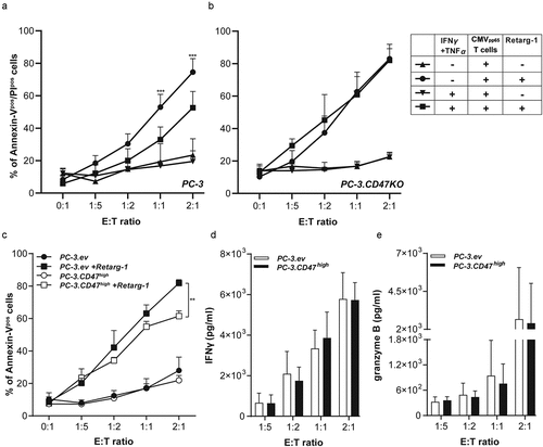
Figure 3. CD47 knockout enhances susceptibility of cancer cells to T cell-mediated cytotoxicity
Cancer cell death (% Annexin-Vpos/PIpos) in (a) parental PC-3 or PC-3.CD47KO (b) FaDu or FaDu.CD47KO cells after co-culturing with PBMCs in the presence of BIS1 at indicated E:T cell ratios for 48 h. (c) The percentage of CD69-expressing CD3pos T cells from data presented in A. (d) IFNγ and Granzyme B levels in co-culture supernatants of A were measured by ELISA. (e) Cell viability of parental FaDu (solid black circles and boxes) and FaDu.CD47KO (open circles and boxes) cells was evaluated for 48 h using the RTCA xCELLigence instrument. After 8 h, PBMCs were added to an E:T cell ratio of 2:1 in the presence (or absence) of BIS1. (f) Cancer cell death in various parental and CD47-KO cell-line pairs was evaluated after treatment as described in A. FaDu (head and neck squamous cell carcinoma); SK-RC-52 (renal cell carcinoma); NCI-H292 (non-small lung cancer); A431 (vulvar squamous cell carcinoma) and OvCAR-3 (ovarian cancer). (g) Cancer cell death in parental FaDu and FaDu.CD47KO cells was evaluated after co-culturing with HLA-matched CMVpp65-specific T cells and target cells in the presence (or absence) of Retarg-1 at indicated E:T cell ratios for 48 h. (h) Caspase-3 activation in FaDu and FaDu.CD47KO cells treated with CMVpp65-specific T cells in the presence of Retarg-1 for 72 h evaluated by the IncuCyte live-cell imaging system. (i) Cancer cell death in various parental and CD47-KO cell-line pairs after treatment as in F. Graphs represent mean ± SD. Experiments in A, B, E, F and H were evaluated by flow cytometry. Statistical analysis in A, E, F and H was performed using unpaired Student’s t‐test with Holm-Sidak post-hoc test.
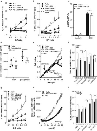
Figure 4. CD47 knockout enhances susceptibility of cancer cells to TRAIL- and FAS-mediated cell death
Induction cancer cell death (% Annexin-Vpos) in various parental and CD47-KO cell-line pairs after treatment with (a) TRAIL or (b) anti-FAS antibody. (c) Cancer cell death in FaDu.CD47KO cells (% Annexin-Vpos/PIpos) after treatment with CMVpp65-specific T cells (E:T cell ratio of 5:1) in the presence of Retarg-1 in combination with either TRAIL-neutralizing antibody mAb 2E5 or FASL-neutralizing antibody mAb 2C101. (d) Evaluation of cell surface expression of CD47 by PC-3 cells transfected with increasing concentrations of CD47-specific siRNA (0.05–5 nM) or control siRNA (% of control siRNA, left y-axis). Evaluation of cancer cell death (% Annexin-Vpos/PIpos, right y-axis) after treatment of CD47-specific siRNA transfected PC-3 cells with (0.1 μg/ml) TRAIL (N = 2). Graphs represent mean ± SD. All experiments were evaluated by flow cytometry. Statistical analyses in A and B were performed using one-way ANOVA with Sidak post-hoc test. Statistical analysis in C was performed using one-way ANOVA with Dunnett post-hoc test.
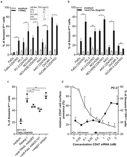
Figure 5. BsAb CD47xEGFR-IgG2s-mediated internalization of CD47 enhances susceptibility of cancer cells to TRAIL-mediated cell death
(a) A431.CD47KO and SK-RC-52.CD47KO cancer cells were transfected with cDNA encoding CD47 C-terminally fused to GFP (CD47-GFP). The percentage of CD47-GFPpos A431 and SK-RC-52 cells was evaluated after treatment with bsAb CD47xEGFR-IgG2s or control antibodies (5 μg/ml) for 48 h. (b) Induction of cancer cell death (% Annexin-Vpos/PIpos) in A431 cells after treatment with bsAb CD47xEGFR-IgG2s, bsAb-controls or indicated anti-CD47 agents (2 μg/ml) for 24 h followed by treatment with TRAIL (1 μg/ml) for 24 h. (c) Induction of cancer cell death in FaDu and FaDu.CD47KO cells was evaluated after treatment with bsAb CD47xEGFR-IgG2s or indicated control antibodies (2 μg/ml) for 24 h, followed by treatment with TRAIL (10 μg/ml) for 24 h. (d) Cancer cell death in various cell lines was evaluated after treatment as in (c) and presented as fold increase compared to medium. (E) A431 cells were treated with increasing concentrations of bsAb CD47xEGFR-IgG2s (0.01–5 μg/ml) for 24 h, after which CD47 cell surface expression (MFI) was evaluated by flow cytometry and compared to untreated cells (left y-axis). Cancer cell death in bsAb CD47xEGFR-IgG2s-treated A431 cells (right y-axis) was evaluated after treatment with TRAIL (1 μg/ml) for 24 h (N = 2). Graphs represent mean ± SD. All experiments were evaluated by flow cytometry. Statistical analysis of A was performed using one-way ANOVA with Dunnett post-hoc test. Statistical analysis in D was performed using unpaired Student’s t-test with Holm-Sidak post-hoc test.
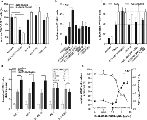
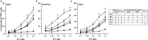
Figure 6. BsAb CD47xEGFR-IgG2s-mediated internalization of CD47 enhances susceptibility of cancer cells to T cell-induced cytotoxicity
Cancer cell death (% Annexin-Vpos/PIpos) in Retarg-1-decorated (a) A431, (b) FaDu or (c) SK-RC-52 target cells evaluated by flow cytometry after treatment with bsAb CD47xEGFR-IgG2s or indicated control antibodies (2 μg/ml) for 24 h followed by treatment with CMVpp65-specific T cells at indicated E:T cell ratios for 24 h. Graphs represent mean ± SD. Statistical analysis was performed using unpaired Student’s t-test with Holm-Sidak post-hoc test.
Supplemental material