
Figures & data
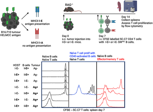
Figure 1. Naïve tumour-specific B cells are poor activators of naïve tumour-specific CD4+ T cells in vivo despite a lymphopenic environment.
(a) Experimental set-up. I-E+ or I-E− RAG−/− host mice were s.c. injected in the flank with 1x10⁶ B16.mHELMCC tumour cells. Mice received I-E+ or I-E− SWHEL B cells and CFSE labelled 5C.C7 CD4+ T cells i.v. 7 days later. (b) Representative flow cytometry plots depicting CD4+ T cell proliferation (CFSE dilution). (c) Frequency and (d) absolute number of 5C.C7 CD4+ T cells in the spleen 7 days after transfer. (e) Representative plots of 5C.C7 CD4+ T cell CFSE dilution and expression of CD62L and CD44. Frequency of (f) effector memory (CD44+CD62L-) and (g) central memory (CD44+CD62L+) 5C.C7 CD4+ T cells in the spleen 7 days post transfer. (h) Representative plots of splenic HEL+ B cells 7 days after transfer. (i) Frequency and (j) absolute number of HEL+ B cells. n=3/group, 12 mice total for experiment. Representative of two independent experiments. ns = not significant, * = p < .05, ** = p < .01,*** = p < .001, **** = p < .0001.
Figure 2. CD40 activated tumour-specific B cells have a limited capacity to activate naïve tumour-specific CD4 T cells.
(a) Experimental set-up. I-E− RAG−/− mice were s.c. injected in the flank with 1x10⁶ B16.mHELMCC tumour cells and after 7 days, naive 5C.C7 CD4+ T cells and activated SWHEL B cells were co-transferred. To generate activated SWHEL B cells, trangenic SWHEL mice were injected10 days prior to transfer with s.c. B16.mHELMCC tumour cells in the flank followed by 2 i.p. injections of anti-CD40 agonistic antibody on days 3 and 6. (b) Representative flow cytometry plots depicting CD4+ T cell proliferation. (c) Frequency and (d) absolute number of 5C.C7 CD4+ T cells in the spleen 7 days after cell transfer. (e) Representative flow cytometric plots of HEL+ B cells in the spleens 7 days after cell transfer. (f) Frequency and (g), absolute number of HEL+ B cells. (h): Mean Fluorescence Intensity (MFI) of MHCII I-E expression on naïve and activated B cells 7 days after adoptive transfer. n = > 4 mice/group, 9 mice total for experiment. ns = not significant, * = p < .05.
Figure 3. Naïve tumour-specific B cells have a limited capacity to activate effector/memory tumour-specific CD4 T cells.
(a) Experimental set-up as for . except that 5C.C7 T cells were harvested from donor mice immunised with antigen emulsified in CFA 21 days prior. (b) Representative flow cytometry plots depicting CD4+ T cell proliferation. (c) Frequency and (d) absolute number of 5C.C7 CD4+ T cells in the spleen 7 days after cell transfer. (e) Representative flow cytometric plots of HEL+ B cells in the spleens 7 days after cell transfer. (f) Frequency and (g), absolute number of HEL+ B cells. n = 3 mice/group, 12 mice total for experiment. Representative of two independent experiments. ns = not significant, * = p < .05, ** = p < .01, **** = p < .0001ss
Supplemental material
Supplemental Material
Download MS Word (304.1 KB)Data availability statement
Raw data were generated at the Centenary Institute. Derived data supporting the findings of this study are available from the corresponding author BF on request.