
Figures & data
Figure 1. S100A10 deficiency prevents lung metastasis.
a) Representative images of B16/F10 lung metastasis in S100A10-deficient or WT mice via luciferase bioluminescence in vivo. b) Representative images of B16/F10 and E0771 lung metastasis nodules in S100A10-deficient or WT mice. c) Quantitation of B16/F10 and E0771 tumor cell focia within the lung tissue (n = 6 individual mice in each group, **p < 0.01). d) Quantitation of B16/F10 and E0771 metastasis lung weight (n = 6 individual mice in each group, **p < 0.01, *p < 0.05). e) Representative images of B16/F10 lung metastasis tissue in WT or KO mice via luciferase bioluminescence and GFP-labeled fluorescence. f) Immunofluorescent analysis S100A10-deficient or WT mice lung sections after GFP-labeled B16/F10 and E0771 inoculation (blue-DAPI, green-GFP, Scale bar, 100 μm). g) The percentage of GFP-positive B16/F10 and E0771 cells in the lung tissue of WT or KO mice detected by flow cytometry (n = 5 individual mice in each group, ***p < 0.001, **p < 0.01). h) Schematic diagram for the lung metastasis of the in situ tumor. i) Representative luciferase bioluminescence images of B16/F10 lung metastasis from in situ S100A10-deficient or WT mice. j) Representative images of B16/F10 lung metastasis tissue from in situ S100A10-deficient or WT mice via luciferase bioluminescence. k) H&E-stained lung sections of S100A10-deficient or WT mice after tail-vein inoculation of B16/F10 cell. l) H&E-stained lung sections of KO or WT mice after subcutaneous injection of B16/F10 cell.
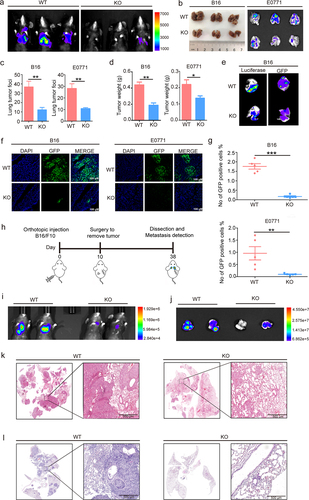
Figure 2. S100A10 deficiency prevents lung pre-metastatic microenvironment formation.
a) Schematic diagram for the tumor-derived exosome induced formation of the pre-metastatic microenvironment. 10 μg of B16/F10 and E0771-derived exosomes in 50 μl of PBS were intravenously injected into the tail vein of S100A10-deficient or WT mice every 2 d. Pre-metastatic microenvironment formation in the lungs of mice after 28 d. b and c) Pro-metastatic gene expression in the lungs of S100A10-deficient or WT mice after B16/F10-derived exosome b) and E0771-derived exosomes c) induction was quantified by qPCR. d and e) Immunofluorescent analysis of lung tissue sections d) and IHC stained lung tissue sections e) of the fibronectin expression of S100A10-deficient mice or WT mice. f) Top 10 biological processes with enrichment of different expression gene sets in S100A10-deficient or WT mice lungs by GO analysis (n = 3 individual mice in each group). g) Top 10 signaling pathways with enrichment of different expression gene sets in S100A10-deficient or WT mice lungs by KEGG analysis (n = 3 individual mice in each group).
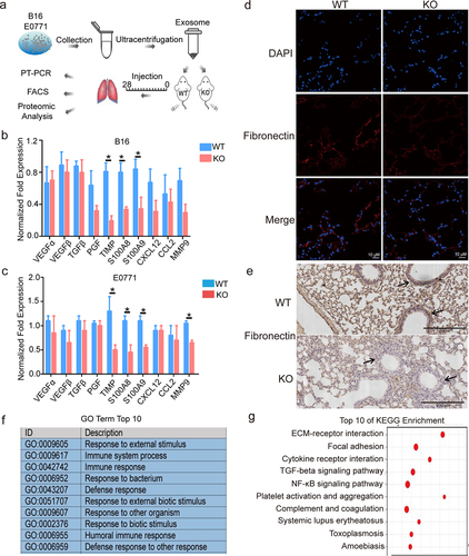
Figure 3. S100A10 deficiency inhibits MDSCs recruitment to the lung pre-metastatic microenvironment.
a) The proportion of MDSCs in the lungs of KO or WT mice after B16/F10 or E0771-derived exosome inoculation was measured via flow cytometry (n = 6 individual mice in each group, *p < 0.05, **p < 0.01). b) The proportion of PMN-MDSCs and M-MDSCs in the lungs of KO or WT mice after B16/F10-derived exosome inoculation was measured via flow cytometry (n = 6 individual mice in each group, *p < 0.05, **p < 0.01). c) The proportion of CD3+CD4+ or CD3+CD8+ T cells in the lungs of KO or WT mice after B16/F10-derived exosome inoculation was measured by flow cytometry (n = 6 individual mice in each group, *p < 0.05, **p < 0.01). d) Immunofluorescent analysis of Gr-1+ cells in WT or KO mice lung sections after B16/F10-derived exosome inoculation (blue-DAPI, green-Gr-1, Scale bar, 25 μm). e) Model diagram of the co-culture system of the T cell, S100A10−/− MDSCs, and S100A10+/+ MDSCs. f) Representative images of CFSE-base proliferation assay of T cells co-cultured with S100A10−/− MDSCs and S100A10+/+ MDSCs by flow cytometry. g) Quantitative T cell proliferation analysis after co-cultured with S100A10−/− MDSCs and S100A10+/+ MDSCs. h) The ARG1, iNOS, and IDO expression of S100A10−/− and S100A10+/+ MDSCs in the lung pre-metastatic microenvironment were quantified by qPCR. i) Anti-Gr-1 Abs or isotype control was injected into the tail-vein of WT and KO mice at 200 µg/mouse the day before the B16/F10LUC were implanted. Subsequently, Gr-1 Abs or isotype control was injected every 3 d into tail veins of WT and KO mice. B16/F10 lung metastasis was monitored via luciferase bioluminescence in vivo.
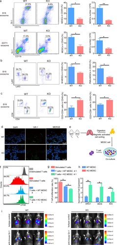
Figure 4. The S100A10 of lung fibroblasts activates CXCL1/CXCL8 expression and increases the migration of myeloid lineage via B16/F10-derived exosome induction.
a) Immunofluorescent analysis of the location of S100A4+ fibroblast and PKH26 marked B16/F10-derived exosome in the lungs of WT mice. b) S100A10 expression in NIH3T3 fibroblast cells stimulated with B16/F10-derived exosomes was detected via immunoblot. c) Chemokine gene expression in NIH3T3 fibroblast cells after B16-derived exosome induction was quantified by qPCR (n = 6 individual cells in each group, *p < 0.05, **p < 0.01). d) Numbers of MDSCs migrating in response to B16/F10 exosome-stimulated fibroblasts conditioned medium (FCM) (n = 6 individual cells in each group, **p < 0.01). e) S100A10 gene knockdown efficiency in NIH3T3 fibroblast cells was detected via immunoblot. f) Chemokine gene expression in S100A10 knockdown NIH3T3 fibroblast cells after B16-derived exosome induction was quantified via qPCR (n = 6 individual cells in each group, *p < 0.05, **p < 0.01). g) Numbers of MDSCs migrating in response to B16/F10 exosome-stimulated S100A10 knockdown NIH3T3 fibroblasts conditioned medium (n = 6 individual cells in each group, **p < 0.01). h) Chemokine gene expression in NIH3T3 fibroblast cells after S100A10 recombinant protein induction was quantified via qPCR (n = 6 individual cells in each group, *p < 0.05, **p < 0.01). i) Numbers of MDSCs migrating in response to the recombinant protein S100A10 stimulated NIH3T3 fibroblasts conditioned medium (n = 6 individual cells in each group, ***p < 0.001).
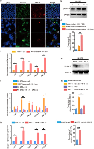
Figure 5. Upregulation of S100A10 expression in lung fibroblasts promotes lung metastasis of tumor in KO mice.
a) The schematic diagram for AAV6-NC or AAV6-S100A10 infection in the lungs of WT and KO mice. Six-week-old WT and KO mice were intratracheally injected with AAV6-NC and AAV6-S100A10. Four weeks after AAV administration, the B16/F10LUC cells were injected into the tail-vein of WT and KO mice. Two weeks after the B16/F10LUC cells were injected, tumor metastasis was assessed via luciferase bioluminescence in vivo. b) qRCR analysis of S100A10 mRNA expression in the lungs of mice from the AAV6-NC and AAV6-S100A10 groups (n = 6 individual cells in each group, ***p < 0.001). c) Western blot analysis of S100A10 expression in the lungs of mice from the AAV6-NC and AAV6-S100A10 groups (n = 3 per group). d) Immunofluorescence staining with S100A4+ fibroblasts and Flag-marked fibroblast-specific AAV6-S100A10 in the lungs of WT mice. e) Statistical analysis of the percent of AAV6-S100A10 transduction in S100A4+ fibroblasts (n = 3 mice, five fields assessed per mouse). f) Representative luciferase bioluminescence images of B16/F10 lung metastasis in WT or KO mice via AAV6-NC and AAV6-S100A10 infection. g and h) The proportion of MDSCs in the lungs of KO or WT mice after AAV6-NC and AAV6-S100A10 infection was measured via flow cytometry (n = 6 individual mice in each group, *p < 0.05, **p < 0.01).
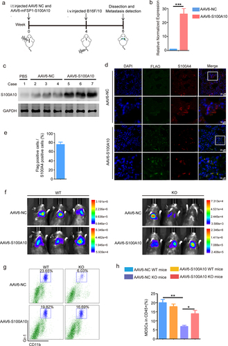
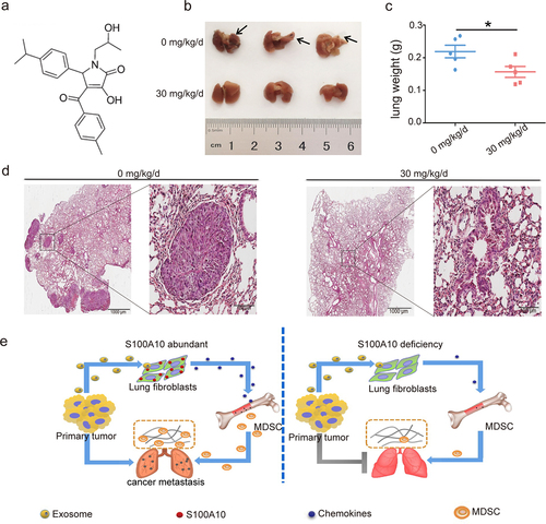
Figure 6. S100A10 inhibitor prevents lung metastasis.
a) Chemical structures of the S100A10 inhibitor 1-substituted 4-aroyl-3-hydroxy-5-phenyl-1 H-pyrrol-2(5 H)-ones. b) Representative images of B16/F10 lung metastasis nodules of C57 mice in treated with vehicle or S100A10 inhibitor 30 mg/kg/d via BFI. c) In vivo quantitation of B16/F10 metastasis lung weight of C57 mice treated with vehicle or S100A10 inhibitor 30 mg/kg/d (n = 5 individual mice in each group, *p < 0.05). d) H&E-stained lung sections of C57 mice treated with vehicle or S100A10 inhibitor 30 mg/kg/d. e) Schematic activation model of S100A10 in host lung fibroblast cells via tumor exosome initiated MDSCs recruitment promotes lung pre-metastatic microenvironment formation and cancer metastasis.
Supplemental material
Supplemental Material
Download MS Word (10.2 MB)Data availability statement
The data that support the findings of this study are available from the corresponding author upon reasonable request.