Figures & data
Figure 1. SDS-PAGE and western blot analysis of Tc24-WT+His in-process samples. Samples were separated on 4–12% Bis-Tris gels under Non-Reduced (A) and Reduced (B) conditions. Lane 1: SeeBlue Plus Molecular Weight marker (10 µl). Lane 2: Starting Material (7.5 µl). Lane 3: IMAC eluate (7.5 µl). Lanes 4–5 QXL eluate (4 µl and 2 µl load). Lanes 6–7: Final Tc24-WT+His (4 µl and 2 µl load). Western blot detection was performed using an in house polyclonal mouse anti-Tc24 antibody at 1:2,500 followed by a goat anti-mouse IgG alkaline phosphatase conjugated secondary antibody at 1:7,500).
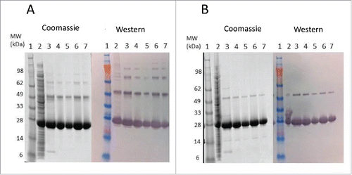
Figure 2. Tc24 amino acid sequence alignment. Amino acid sequence alignment of wild-type (Tc24-WT) and the cysteine-mutated Tc24 constructs (Tc24-C2 and Tc24-C4). Cysteine residues are highlighted in red and mutated serine residues in green.
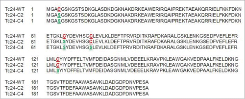
Figure 3. Western blot comparison of Tc24-WT (A), Tc24-C2 (B), and Tc24-C4 (C) purified proteins. Lanes 1–3: Non-Reduced. Lane 4: SeeBlue Plus Molecular Weight Marker. Lanes 5–7 Reduced. Lanes 1 and 5: Sample before size-exclusion chromatography (SEC) 8 µg load. Lanes 2 and 6: Post SEC low load (3 µg). Lanes 3 and 7: Post SEC high load (8 µg). Detection was performed using mouse polyclonal antibody against Tc24 expressed in Pichia pastoris as primary antibody diluted 1:2,500 in PBST and an alkaline phosphatase conjugated goat anti-mouse secondary antibody diluted 1:7,500 in PBST.
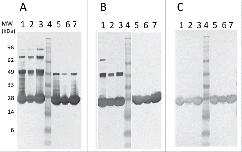
Table 1. Comparison of yield and purity for Tc24 constructs.
Figure 4. SDS-PAGE analysis of in-process and final Tc24-C4 protein samples. Protein samples were separated on a 4–12% Bis-Tris SDS PAGE gel under reducing (A) and non-reducing (B) conditions. Lanes 1: SeeBlue Plus 2 molcular weight marker. Lanes 2: Cell Lysate (3 µg). Lane 3: QXL1 elution (3 µg). Lane 4: QXL2 elution (3 µg). Lane 5: Concentrated QXL (8 µg). Lane 6: SEC elution (3 µg). Lane 7: SEC elution (8 µg).
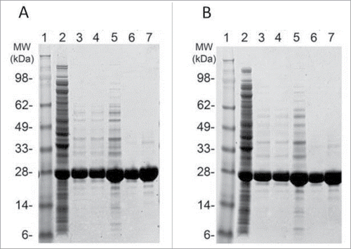
Figure 5. Stability assessment of Tc24. Western blot of different Tc24 constructs, taken after storing the proteins for 10 d at 4°C in PBS. Lane 1: Tc24-WT+His, Lane 2:Tc24-WT+His, alkylated, Lane 3: Tc24-WT, Lane 4: Tc24-C2, Lane 5: Tc24-C4. A Ponceau-stained Mark 12 protein ladder was used as a MW reference. Detection was performed using mouse polyclonal antibody against Tc24 expressed in Pichia pastoris as primary antibody diluted 1:2,500 in PBST and an alkaline phosphatase conjugated goat anti-mouse secondary antibody diluted 1:7,500 in PBST.
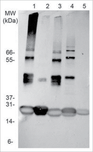
Figure 6. Hydrodynamic radius and polydispersity of Tc24 antigens after 3 d at 4°C. Tc24-C4 was noticeably the most monodispersed product.
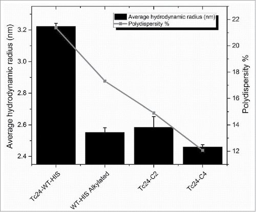
Figure 7. Structural comparison of Tc24 constructs. A) Circular Dichroism (CD). Far UV CD spectrum of different constructs of Tc24 were taken on a Jasco J-1500. All tested Tc24 protein have a virtual identical CD profile with overlapping spectra. Negative peaks at 222 nm and 208 nm and a positive peak at 193 nm indicate that Tc24 is an α-helical protein. B) Thermal melting profile of Tc24-WT and Tc24-C4 measured using Protein Thermal Shift™ kit (Life Technologies).
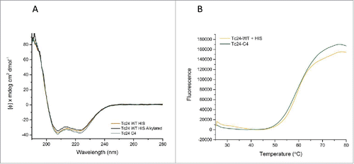
Figure 8. IFNγ measurements after homologous or heterologous stimulation of vaccinated mice by ELISA. IFNγ concentrations in supernatants from splenocytes harvested from mice vaccinated with the indicated protein combined with E6020 emulsified in AddaVaxTM and re-stimulated in vitro with homologous recombinant protein (A) or mice vaccinated with Tc24-WT No His combined with E6020 emulsified with AddaVaxTM and re-stimulated in vitro with heterologous protein as indicated (B). No statistically significant differences in antigen specific IFNγ secretion were observed. (C) IFNγ secretion from mice vaccinated with Tc24 C4 combined with E6020 in a stable squalene emulsion was significantly increased compared to mice vaccinated with Tc24 C4 (protein only) or E6020 SE alone (adjuvant only control).
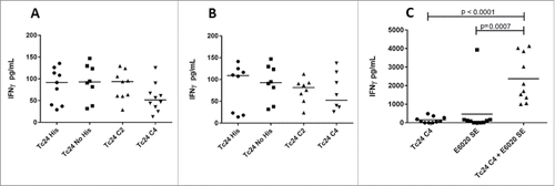
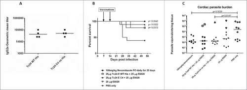
Figure 9. Measurement of IgG2a in vaccinated mice by ELISA. Anti-Tc24 IgG2a antibody titers in terminal serum were measured by ELISA for wild-type and mutant Tc24 constructs. Antibody titers in mice vaccinated with Tc24-C2 no His or Tc24-C4 no His were not significantly different from mice vaccinated with Tc24-WT no His.
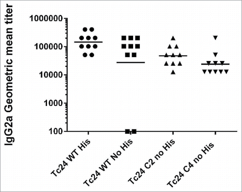
Figure 10. Specificity of antibodies from infected mice against wild-type and mutant Tc24 constructs, survival and tissue parasite burdens of acutely infected mice vaccinated therapeutically. Antibodies from mice infected with T. cruzi parasites were evaluated for specificity against either Tc24–WT (His-tagged) or Tc24-C4 by ELISA (Panel A). Plates were coated with either antigen and bound antibodies were detected using labeled goat anti-mouse IgG2a secondary antibodies. Geometric mean titers were calculated. Acutely infected mice were vaccinated therapeutically with either Tc24-WT (His-tagged) or Tc24-C4 combined with E6020 SE. Survival was monitored daily (Panel B) and tissue parasite burdens were measured by quantitative PCR from cardiac tissue (Panel C). Group 1 (●) treated with benznidazole; Group 2 (□) vaccinated with Tc24-WT His + E6020 SE; Group 3 (▲) vaccinated with Tc24-C4 + E6020 SE; Group 4 (▽) vaccinated with E6020 SE; Group 5 (♦) vaccinated with PBS only. Open symbols in panel C indicate mice that did not survive until the end of the study.