Figures & data
Figure 1. The morphology of mature DCs After stimulated with TNF-α, HPVm16E7-pulsed DCs in irregular shape showed mature characteristics of larger soma with plenty of dendritic long and small bulges on their surfaces (400 × magnification). White arrow indicates long bulge and black arrow indicates small bulge.
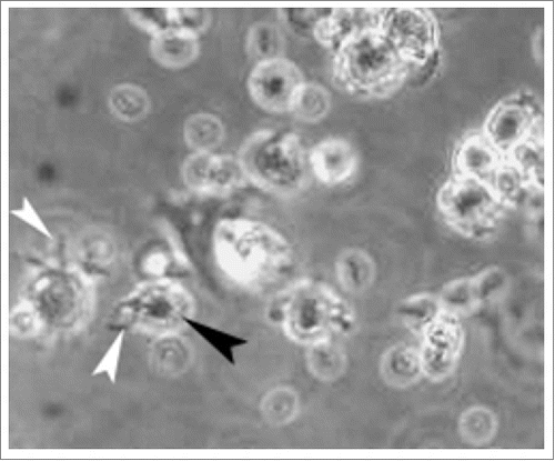
Table 1. Percentage of DCs expressing CD1 a, CD80, CD83, CD86, HLA-DR.
Figure 2. Cytotoxicity assay (A) Detected by flow cytometry, 23.6% CaSki cells expressed PDL1. (B)The cytotoxicity of effector cells at 10:1, 30:1 and 90:1 E/T ratios against CaSki cells was analyzed using CCK 8 assay. When lymphocytes incubated with MPDL3280A, DCCIKs increased the cytotoxic activity against CaSki cells with 37.9% at the E:T of 30 in relative to DCCIKs in the absence of MPDL3280A (p < 0 .01).
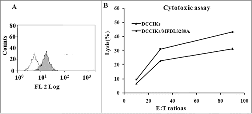
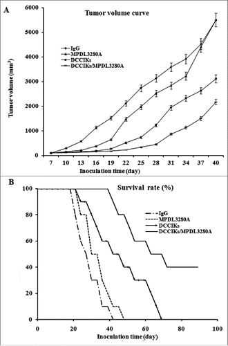
Figure 3. In vivo antitumor activity analysis. (A) The therapeutic effect of DCCIKs in combination with MPDL3280A against mice bearing tumors was investigated in the groups of 10 BALB/C mice received 1×10⁶ CaSki cells. The therapy was initiated with normal human IgG, DCCIKs, MPDL3280A and DCCIKs in combination with MPDL3280A on day 7. Tumor volume and time experiment was analyzed using student's t-test. *p < 0.01(DCCIKs/MPDL3280A vs DCCIKs or MPDL3280A or IgG control); <0.05 (DCCIKs vs MPDL3280A or IgG control) (B) Survival rates were analyzed using the Kaplan-Meier method (log-rank test). The experiments were repeated, producing a significant statistical difference. *p < 0.01(DCCIKs/MPDL3280A vs DCCIKs or MPDL3280A or IgG control); <0.05 (DCCIKs vs MPDL3280A or IgG control).