Figures & data
Table 1. Primer sequence
Figure 1. Elevated circPIP5K1A and JAK1 but reduced miR-552-3p are presented in the serum sample of T2DM patients.
A: RT-qPCR to detect circPIP5K1A, miR-552-3p, and JAK1 in the serum of T2DM patients, n = 58; ***P < 0.001; B: The analysis of liner link between circPIP5K1A and miR-552-3p, miR-552-3p and JAK1, circPIP5K1A and JAK1 in T2DM serum was carried out by Spearman’s correlation coefficient.
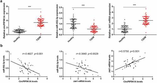
Figure 2. Downregulated circPIP5K1A restrains the inflammation, oxidative damage, and apoptosis of INS-1 β cell induced via glycolipid toxicity.
A/B: RT-qPCR to detect circPIP5K1A; C: CCK-8 to detect INS-1E cell viability and each bar represent the mean ± standard deviation of three independent experiments; D, E: Flow cytometry to detect INS-1E cell apoptosis and measure cellular ROS. Each bar represented the mean ± standard deviation of three independent experiments in which *value show significant difference as compared to the control (*P < 0.05). F, G: ELISA to detect inflammatory factors TNF-α and IL-6 in INS-1E cells and measure secreted insulin. Cells were transfected with 50 nmol of oligonucleotides and incubated for 48 hours before subsequent assays. *P < 0.05, **P < 0.01, ***P < 0.001.
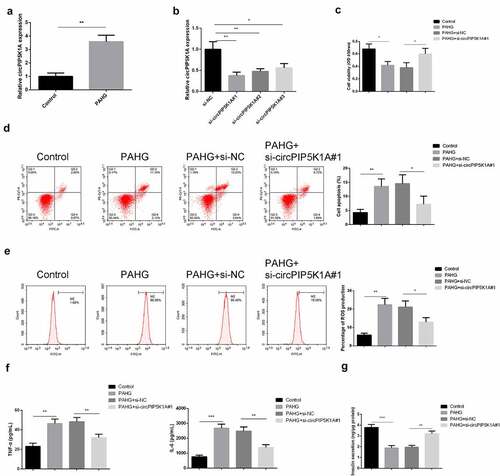
Figure 3. CircPIP5K1A adsorbs miR-552-3p in INS-1E cells.
A: Nuclear and cytoplasmic separation experiments to testify circPIP5K1A was mainly situated in the cytoplasm; B: Bioinformatics website starbase to forecast the binding site of circPIP5K1A and miR-552-3p; C: Dual luciferase verification of circPIP5K1A and miR-552-3p’s binding; D, E: RNA pull-down to examine the enrichment of miR-552-3p on circPIP5K1A and miR-552-3p after depressive circPIP5K1A. Cells were transfected with 50 nmol of oligonucleotides and incubated for 48 hours before subsequent assays. Graph presented mean ± standard deviation results of three independent experiment *P < 0.05, **P < 0.01, ***P < 0.001.
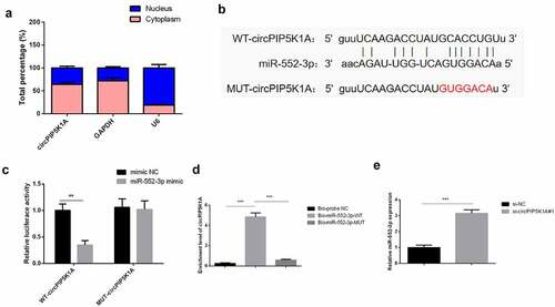
Figure 4. Downregulating miR-552-3p turns around that dissipative circPIP5K1A reduces the toxic effect of glycolipid toxicity on INS-1 β cell.
A: RT-qPCR to test circPIP5K1A and miR-552-3p; B: CCK-8 to examine INS-1E cell viability; C, D: Flow cytometry to detect INS-1E cell apoptosis and measure cellular ROS. Each bar represented the mean ± standard deviation of three independent experiments in which *value show significant difference as compared to the control (*P < 0.05). E, F: ELISA to detect inflammatory factors of TNF-α and IL-6 in INS-1E cells and measure secreted insulin. Cells were transfected with 50 nmol of oligonucleotides and incubated for 48 hours before subsequent assays. Graph presented the mean ± standard deviation of three independent experiments in which *value show significant difference as compared to the control (*P < 0.05) *P < 0.05, **P < 0.01, ***P < 0.001.
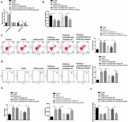
Figure 5. JAK1 acts as a targeted mRNA of miR-552-3p.
A: Bioinformatics website starbase to forecast the binding site of miR-552-3p and JAK1; B, C: Dual luciferase verification and RIP detection of miR-552-3p and JAK1’s binding; D, E: RT-qPCR and WB to detect JAK1 after elevated or depressive miR-552-3p. Cells were transfected with 50 nmol of oligonucleotides and incubated for 48 hours before subsequent assays. Graph presented the mean ± standard deviation of three independent experiments in which *value show significant difference as compared to the control (*P < 0.05) *P < 0.05, **P < 0.01, ***P < 0.001.
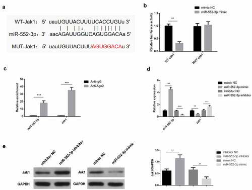
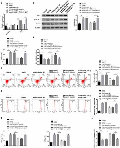
Figure 6. MiR-552-3p refrains the inflammation, oxidative damage, and apoptosis of glycolipid toxicity-stimulated INS-1 β cells via targeting the JAK1-STAT3 pathway.
A, B: RT-qPCR and WB to detect miR-552-3p, JAK1, p-STAT3, STAT3; C: CCK-8 to detect INS-1E cell viability; D, E: Flow cytometry to detect INS-1E cell apoptosis and measure cellular ROS; F, G: ELISA to detect inflammatory factors TNF-α and IL-6 in INS-1E cells and measure secreted insulin. Cells were transfected with 50 nmol of oligonucleotides and incubated for 48 hours before subsequent assays. Graph presented the mean ± standard deviation of three independent experiments in which *value show significant difference as compared to the control (*P < 0.05) *P < 0.05, **P < 0.01, ***P < 0.001.