Figures & data

Figure 1. SHMT2 is up-regulated in OSCC cells. (a-b) The detection of SHMT2 mRNA and protein levels employed RT-qPCR and western blot in OSCC cell lines. *P < 0.05, ***P < 0.001 vs. HOK.
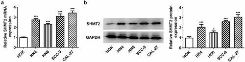
Figure 2. SHMT2 interference ameliorates the initiation of OSCC. (a-b) With the aid of RT-qPCR and western blot, the knockdown efficiency of SHMT2 was determined. (c-d) CCK-8 and colony formation assays appraised cell proliferation. (e-f) The relative apoptosis rate was estimated with the employment of TUNEL. (g-h) The expression of proteins linked to apoptosis was examined with the application of western blot. ***P < 0.001 vs. si-NC.
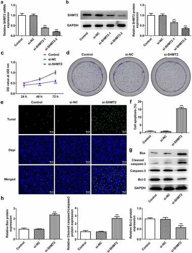
Figure 3. SHMT2 deficiency suppresses the progression of OSCC. (a-b) Wound healing and transwell assays appraised cell migration and invasion. (c) Western blot analysis of MMP2 and MMP9 expression. (d) Western blot analysis of the expression of EMT-related factors. *P < 0.05, **P < 0.01, ***P < 0.001 vs. si-NC.
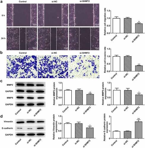
Figure 4. ILF2 binds to SHMT2 and can be downregulated by SHMT2 silencing in OSCC cells. (a-b) The interaction between SHMT2 and ILF2 was found through the MINT and BioGRID databases. (c) ILF2 level in oral tumor tissue was analyzed using TNMplot database. (d-e) RT-qPCR and western blot analysis of ILF2 expression. **P < 0.01, ***P < 0.001 vs. HOK. (f) ILF2 expression in SHMT2-knockdown CAL-27 cells was detected with the use of western blot. ***P < 0.001 vs. si-NC. (g) The interaction of SHMT2 and ILF2 was identified using Co-IP.
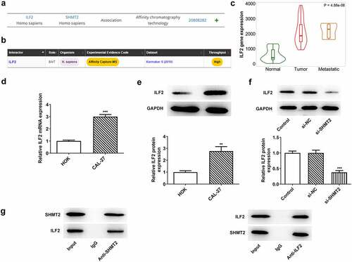
Figure 5. ILF2 upregulation reverses the impacts of SHMT2 knockdown on the occurrence of OSCC. (a-b) RT-qPCR and western blot analysis of the overexpression efficiency of ILF2. ***P < 0.001 vs. Oe-NC. (c) CCK-8 assay appraised cell viability in CAL-27 cells transfected with si-SHMT2 and Oe-ILF2. (d) Cell proliferation was identified utilizing colony formation assay in CAL-27 cells transfected with si-SHMT2 and Oe-ILF2. (e-f) TUNEL assay appraised cell apoptosis. (g-h) Western blot analysis of the expression of apoptosis-related factors. **P < 0.01, ***P < 0.001 vs. si-NC; #P < 0.05, ##P < 0.01, ###P < 0.001 vs. si-SHMT2+ Oe-NC.
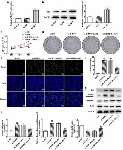
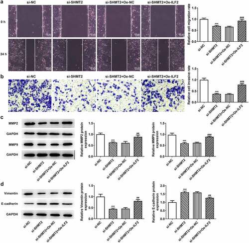
Figure 6. ILF2 elevation restores the impacts of SHMT2 knockdown on OSCC cell migration, invasion and EMT. (a-b) Wound healing and transwell assays appraised cell migration and invasion. (c) Western blot analysis of MMP2 and MMP9 expression. (d) Western blot analysis of the expression of EMT-related factors. ***P < 0.001 vs. si-NC; ##P < 0.01, ###P < 0.001 vs. si-SHMT2+ Oe-NC.