
Figures & data

Table 1. Primers
Figure 1. The identification of BMSCs-derived exosomes. (a) The morphology of BMSCs by an inverted microscope. (b) Flow cytometry analysis of the BMSCs surface markers. (c) Exosomes were isolated from BMSCs under transmission electron microscopy (TEM) identification. (d) Western blot analysis of the exosome surface markers.
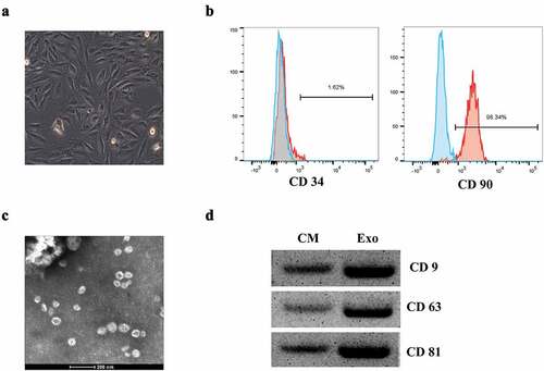
Figure 2. BMSCs-derived exosomes (Exos) enhanced the activities of hFOB1.19 cells. (a) hFOB1.19 cell proliferation determined by Cell counting kit-8 (CCK-8) assay after treated with BMSCs-derived exosomes at different time point. (b) Osteoblastic differentiation in hFOB1.19 cells evaluated by Alizarin Red staining after treated with BMSCs-derived exosomes. (c) Alkaline phosphatase (ALP) staining of hFOB1.19 cells after treated with BMSCs-derived exosomes. (d) hFOB1.19 cells cycle detected by flow cytometry after treated with BMSCs-derived exosomes. * p < 0.05, ** p < 0.01, *** p < 0.001, and NS indicates no significant difference.
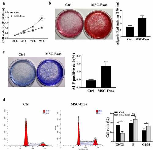
Figure 3. BMSCs-derived exosomes (Exos) enhanced the activities of hFOB1.19 cells via exosomal miR-21-5p. (a) Relative level of miR-21-5p in BMSCs-derived exosomes after transfection. (b) hFOB1.19 cell proliferation determined by Cell counting kit-8 (CCK-8) assay after treated with BMSCs-derived exosomes with changed miR-21-5p. (c) Alkaline phosphatase (ALP) staining of hFOB1.19 cells after treated with BMSCs-derived exosomes with changed miR-21-5p. (d) Osteoblastic differentiation of hFOB1.19 cells evaluated by Alizarin Red staining after treated with BMSCs-derived exosomes with upregulated or downregulated miR-21-5p. ** p < 0.01, *** p < 0.001.
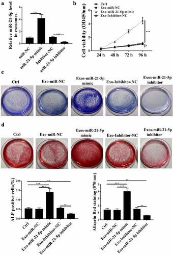
Figure 4. Kruppel-like factor 3 (KLF3) was targeted by miR-21-5p. (a) Relative level of miR-21-5p in hFOB1.19 cells after miR-21-5p mimics transfection. (b) The putative interaction predicted by Targetscan. (c) The impacts of miR-21-5p mimics on WT/MUT KLF3 in hFOB1.19 cells by luciferase reporter assay. (d and e) Relative level of KLF3 in hFOB1.19 cells after transfection of miR mimics or inhibitor determined by qRT-PCR (d) and western blot (e). * p < 0.05, ** p < 0.01, *** p < 0.001.
Figure 5. KLF3 mediated the impacts of miR-21-5p on the activities of hFOB1.19 cells. (a and b) Relative mRNA (a) and protein (b) expression level of KLF3 in hFOB1.19 cells after treating with BMSCs-Exo sh-KLF3. (c and d) Alkaline phosphatase (ALP) staining (c) and osteoblastic differentiation (d) of hFOB1.19 cells after co-treated with BMSCs-derived exosomes (Exos) carrying miR-21-5p inhibitor and sh-KLF3. * p < 0.05, ** p < 0.01, *** p < 0.001.
Supplemental Material
Download Zip (9.1 MB)Data availability statement
The data that support the findings of this study are available from the corresponding author by reasonable request.