
Figures & data

Figure 1. DXP decreases the expressions of inflammatory cytokines in the serum of CLP-induced mice model. (a-d) Expressions of TNF-α, IL-1β, IL-6 and MCP-1 in the serum of mice was detected by ELISA. ***P < 0.001 vs Control; ###P < 0.001 vs CLP.
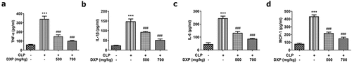
Figure 2. DXP reduces CLP-induced damage of kidney function, inflammation and pathological tissue in mice. (a-b) Biochemical kits were used to detect the expressions of kidney function indicators BUN and Cre in the serum of mice. (c) Expressions of NGAL and Kim1 in kidney tissues was assayed adopting western blot. (d) Expressions of TNF-α, IL-1β, IL-6 and MCP-1 in kidney tissues of mice was examined applying western blot. (e) Kidney histopathological damage was observed using HE staining. ***P < 0.001 vs Control; #P < 0.05, ##P < 0.01, ###P < 0.001 vs CLP.
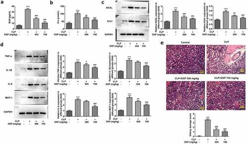
Figure 3. DXP reduces CLP-induced apoptosis in the kidney tissue of mice. (a) Level of apoptosis in the kidney tissues of mice was observed using TUNEL staining. (b-c) Expressions of apoptosis-related proteins in kidney tissues of mice were examined with the application of western blot. ***P < 0.001 vs Control; ###P < 0.001 vs CLP.
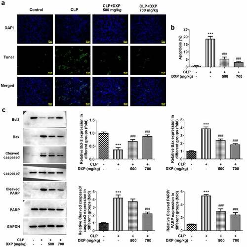
Figure 4. DXP reduces CLP-induced damage of liver function, inflammation, antioxidant enzymes and pathological tissue of mice. (a-c) Biochemical kits were used to assay liver function impairment indicators ALT, AST, ALP. (d) Assay of MPO activity in the liver of mice was undertaken using MPO kit. (e) Expressions of inflammatory factors TNF-α, IL-1β, IL-6, and MCP-1 in the liver tissues of mice were tested employing western blot. (f-g) Expressions of GSH and SOD in liver tissues of mice were measured using GSH and SOD kits. (h-i) Observation of liver histopathological damage in mice was conducted adopting HE staining. ***P < 0.001 vs Control; #P < 0.05, ##P < 0.01, ###P < 0.001 vs CLP.

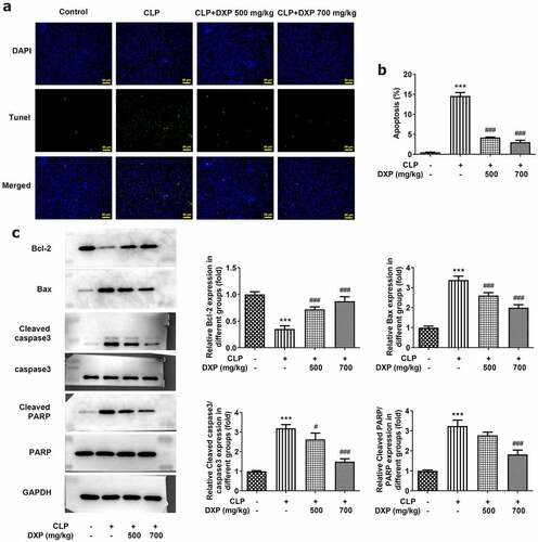
Figure 5. DXP reduces CLP-induced apoptosis in the liver of mice. (a) Measurement of apoptosis level in the hepatocytes of mice was carried out using TUNEL staining. (b-c) Detection of apoptosis-related protein expressions in the liver tissues of mice through western blot. ***P < 0.001 vs Control; #P < 0.05, ###P < 0.001 vs CLP.
Data availability statement
All data generated or analyzed during this study are included in this article.