Figures & data
Figure 1. Expression of miR106a and Connexin43 in SNHL patients. MiR-106a was significantly upregulated, connexin 43 was reduced in SNHL patients. The level of miR-106a in the peripheral blood of SNHL patients and health subjects was determined by RT-PCR. The connexin43 was detected by ELISA. (*p < 0.05, **p < 0.01 vs. Health).
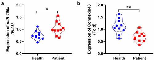
Figure 2. The isolated rat cochlear MCs were identified and the incubation concentration of GO was determined. A. The isolated rat cochlear MCs were identified by checking the expression of CK18 using the immunofluorescence assay. B. The OD value was measured using the CCK-8 assay (**p < 0.01, ***p < 0.001). Scale bar: 50 μm.
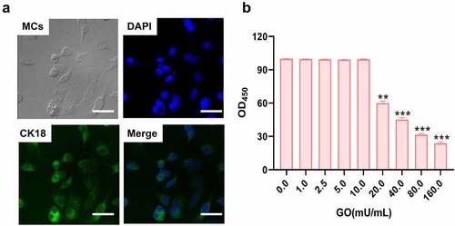
Figure 3. The apoptosis and oxidative stress in GO-treated MCs were ameliorated by the knockdown of miR-106a. MCs were treated with 20 mU/mL GO with or without inhibitor NC and miR-106a inhibitor, respectively. A. The OD value was checked utilizing the CCK-8 assay. B. The apoptotic rate was investigated by the flow cytometry assay. C. The level of Bcl-2, Bax, and caspase-3 was evaluated by the Western blotting assay. D. The DCFH-DA assay was utilized to determine the ROS level in MCs. E. The production of MDA was investigated utilizing the commercial kit. F. The production of SOD was investigated utilizing the commercial kit. G. The level of Nrf2, Keap1, and HO-1 was confirmed by the Western blotting assay (**p < 0.01 vs. control, ##p < 0.01 vs. GO+ inhibitor NC).
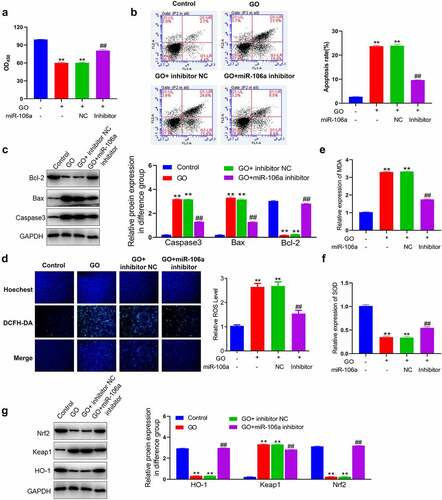
Figure 4. MiR-106a mediated connexin-43 by targeting the 3ʹUTR region and A. The level of miR-106a was measured using the RT-PCR assay. B. The expression level of connexin-43 was investigated by the Western blotting assay (**p < 0.01 vs. control, ##p < 0.01 vs. GO+ inhibitor NC). C. The correlation between miR-106a and connexin-43 was predicted and confirmed using the dual luciferase gene reporter assay (**p < 0.01 vs. miR-NC).
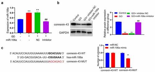
Figure 5. Knockdown of connexin-43 abolished the inhibitory property of miR-106a inhibitor against GO-induced apoptosis and oxidative stress in MCs. MCs were treated with 20 mU/mL GO and miR-106a inhibitor with or without a siRNA targeting connexin-43. A. The OD value was checked utilizing the CCK-8 assay. B. The apoptotic rate was investigated by the flow cytometry assay. C. The level of Bcl-2, Bax, and caspase-3 was evaluated by the Western blotting assay. D. The DCFH-DA assay was utilized to determine the ROS level in MCs. E. The production of MDA was investigated utilizing the commercial kit. F. The production of SOD was investigated utilizing the commercial kit. G. The level of Nrf2, Keap1, and HO-1 was confirmed by the Western blotting assay (**p < 0.01 vs. GO+ inhibitor NC+ siR-NC, #p < 0.05 vs. GO+ miR-106a inhibitor, ##p < 0.01 vs. GO+ miR-106a inhibitor).
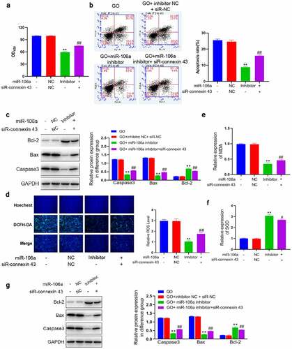
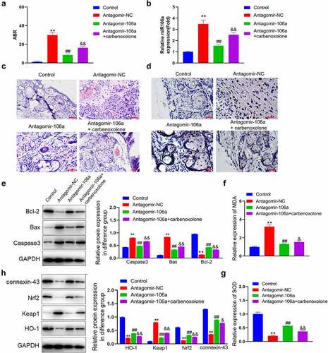
Figure 6. Pathological state and the level of oxidative stress in SNHL rats were alleviated by antagomir-106a. SNHL rats were administered with antagomir-NC, antagomir-106a, or antagomir-106a combined with carbenoxolone, respectively. A. The ABR values were recorded. B. The level of miRNA106a in SNHL rats was detected by RT-PCR. C. The pathological changes in cochlea tissues were determined by HE staining. D. The apoptosis in cochlea tissues was evaluated by the TUNEL assay. E. The level of Bcl-2, Bax, and caspase-3 was evaluated by the Western blotting assay. F. The concentration of MDA was measured using the commercial kit. G. The concentration of SOD was measured using the commercial kit.H. The expression level of Nrf2, Keap1, and HO-1 was evaluated by the Western blotting assay (**p < 0.01 vs. control, ##p < 0.01 vs. antagomir-NC, & p < 0.05 vs. antagomir-106a, && p < 0.01 vs. antagomir-106a). Scale bar: 100 μm.