Figures & data
Table 1. qPCR primer sequences
Figure 1. Icariin improves cardiac function after myocardial infarction: (a) Thoracic echocardiography of mice after 28 days of treatment compared to the sham group. (b) Illustration of H&E staining and measurement of the cross-sectional area of the fibers in the hearts (× 100). (c) Digitalized slides of Masson’s trichrome-stained heart paraffin slices and collagen volume. Collagen has been dyed in blue, and the cytoplasm has been stained in red, respectively (× 100).
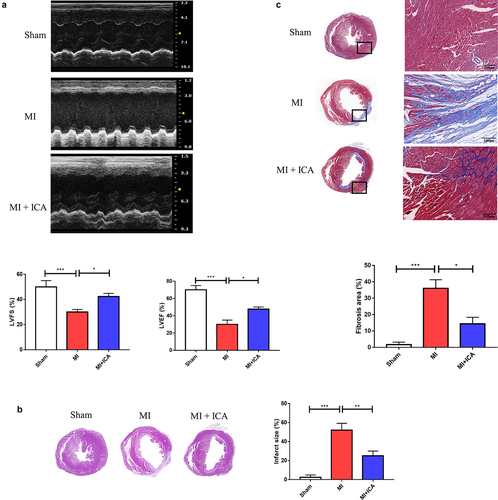
Figure 2. Icariin prevents myocardial cell apoptosis: (a) Mice myocardial were stained with TUNEL, *P < 0.05. (b) The protein expression of Bcl-2, Bax, cytochrome c, cleaved caspase-3, and GAPDH (control) were determined by Western blotting. Each experience was repeated 3 times *P < 0.05.
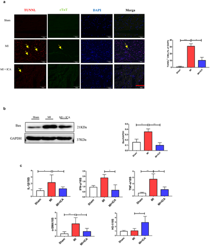
Figure 3. Icariin improves mice immune system: Flow cytometric analysis was performed to assess the population of lymphocytes (CD8+ CTL cells and CD3+ CD4++IFN-γ+ Th1) in the spleen and heart.
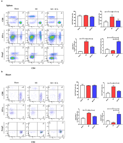
Figure 4. Icariin promotes activation of Nrf2/HO-1 by alleviating inflammatory factors. Signaling pathways: (a) Immunohistochemical was performed to check the expression of IL-17 (× 100); the experiment was repeated 3 times with means *P < 0.05. (b) qPCR was performed to evaluate the mRNA expression of IL1β, HO-1, TNF-α, and α-SMA compared to 18S. Each experience was repeated 3 times *P < 0.05. (c) The protein expression of HO-1, Nrf2, and GAPDH (control) was determined by Western blotting. Each experience was repeated 3 times *P < 0.05.
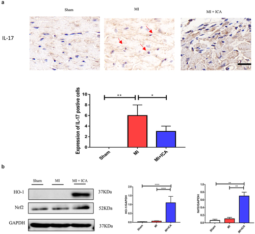
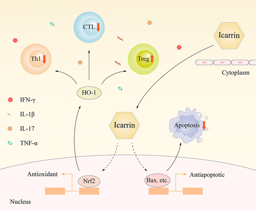
Figure 5. Molecular mechanism by which Icariin induces myocardial cell protection.