
Figures & data
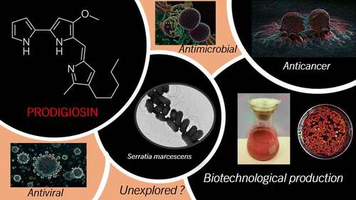
Figure 1. Comparative chemical structure of prodigiosin and its different isoforms (with permission from [Citation18]).
![Figure 1. Comparative chemical structure of prodigiosin and its different isoforms (with permission from [Citation18]).](/cms/asset/10aad70c-5a5b-4699-91d3-363c05348bc8/kbie_a_2084498_f0001_b.gif)
Table 1. Summary of PG production reported in the literature.
Figure 2. Schematic steps for Prodigiosin purification.
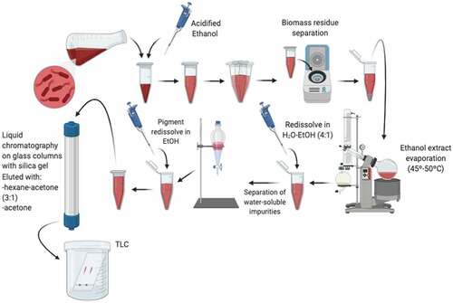
Table 2. Antibacterial activity of Prodigiosin.
Table 3. Antifungal activity of Prodigiosin.
Table 4. Anticancer activity of Prodigiosin.
Figure 3. Obatoclax mesylate, a derivative of prodigiosin with high anticancer activity.
Table 5. Different drug delivery systems for prodigiosin vehiculation.
Figure 4. Schematic process of the prodigiosin nanomicelles production (modified from Gong et al.[Citation18]).
![Figure 4. Schematic process of the prodigiosin nanomicelles production (modified from Gong et al.[Citation18]).](/cms/asset/a35cb714-8e3c-4192-9c1a-b9a8f8ba9fb6/kbie_a_2084498_f0004_oc.jpg)
Figure 5. SEM micrograph showing the morphologies of (a) PLGA/Ge-PG electrospun scaffold and (b) PLGA/Ge-F127/PG scaffold (with permission from [Citation117]).
![Figure 5. SEM micrograph showing the morphologies of (a) PLGA/Ge-PG electrospun scaffold and (b) PLGA/Ge-F127/PG scaffold (with permission from [Citation117]).](/cms/asset/45da5e25-5842-4b44-8f33-48edbe306e25/kbie_a_2084498_f0005_oc.jpg)
Figure 6. The potential application of 3D printing to design aortic root starting from the computed tomography cross-sectional view, followed by the 3D computational Model to finally obtain a 3D-Printed phantom (with permission from [Citation121]).
![Figure 6. The potential application of 3D printing to design aortic root starting from the computed tomography cross-sectional view, followed by the 3D computational Model to finally obtain a 3D-Printed phantom (with permission from [Citation121]).](/cms/asset/08f88377-0a51-47a4-97c1-136916da3dcc/kbie_a_2084498_f0006_oc.jpg)