Figures & data
Figure 1. Neutrophil recruitment. Neutrophils are derived from hematopoietic stem cells of the bone marrow and developed in the bone marrow. The sequential steps of classical neutrophil recruitment from the vascular system to tissues in response to infection are shown. Neutrophils undergo integrin-mediated crawling to migrate. The migration of endothelial cell junctions is mediated by several other adhesion molecules. The release of proteolytic enzymes allows neutrophils to enter the extravascular spaces. Extravascular neutrophils follow a chemotactic gradient to reach the tumor site. Neutrophils are involved in various stages of tumorigenesis and development, including tumorigenesis, proliferation, and metastasis.
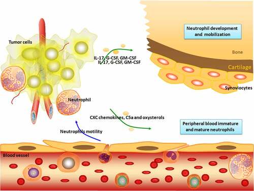
Figure 2. Schematic illustration of the procedure of PET tracer based molecular imaging. After producing a suitable PET tracer and injecting the radioactive tracer, positrons were emitted into the body of the subject, and PET was detected and imaged. 18F-FDG is similar to glucose metabolism in cells and is currently the largest imaging agent available for clinical use. Phosphorylated glucose (glucose-6 P) can be further metabolized by cells, while phosphorylated 18F-FDG cannot be further metabolized, so it is considered ‘trapped.’ 18F-FDG PET/CT metabolic parameters have a good correlation with the clinical prognostic indicator NLR, suggesting that it may be more advantageous than traditional indicators for evaluating tumor prognosis.
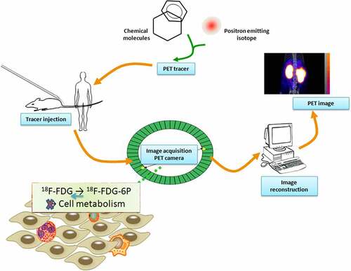
Table 1. Nanoparticle-remodeled neutrophils.
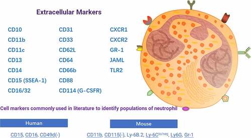
Figure 3. Schematic diagram of neutrophil-specific markers. Intratumoral neutrophils and their surface markers can be used as targets for imaging diagnostic techniques to dynamically monitor tumor progression and inflammation in the microenvironment.