Figures & data
Figure 1 HGF reduces sorafenib anti-proliferative effects in HCC cells. HepG2 and Huh7 cells were treated with or without 50 ng⁄ml HGF for 24 h, then treated with indicated compounds for 72 h. Each point, the mean ± SD for three independent experiments. The IC50 was calculated by non-linear regression analysis using GraphPad Prism software.
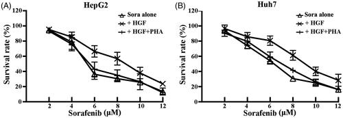
Figure 2 HGF protects HCC cells from sorafenib-induced apoptosis. Apoptosis of HepG2 and Huh7 cells detected by the Annexin V–FITC/PI assay. Cells were treated with or without 50 ng/ml HGF for 24 h, thentreated with indicated compounds for 48 h. The rate of apoptosis was determined using a flow cytometer, and data were analyzed using Kaluza software and were reported as the mean ± SD. The results are representative of three independent experiments. p<.05.
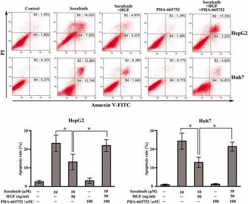
Figure 3 HGF antagonizes sorafenib anti-metastatic effect in HCC cells. HepG2 and Huh7 cells were treated with sorafenib (5 μM) alone or HGF (50 ng/ml) and sorafenib. The cells were then incubated for 24 h (migration assay) or 48 h (invasion assay). (A) Migration abilities were measured using a transwell assay without Matrigel. (B) Invasion abilities were measured using a transwell assay with Matrigel. Data shown represent the mean ± SD. p<.05.
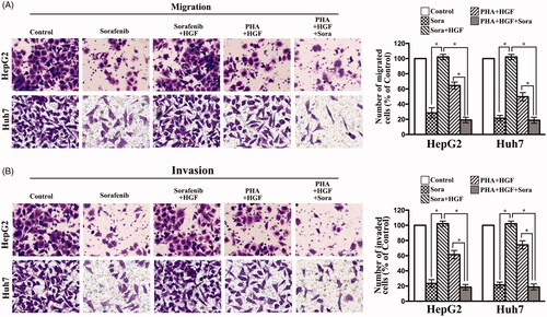
Figure 4 HGF rescue HCC cells through MET-dependent activation of AKT and ERK1/2 pathways. HepG2 and Huh7 cells were starved in serum-reduced (1% FBS) media for 12 h before adding either sorafenib (5 μM) or PHA-665752 (100 nM). After incubation for 6 h and stimulation with 50 ng/mL HGF for 10 min, cells were lysed for Western blot analysis. Independent experiments were performed at least three times, and the results from a representative experiment are shown.
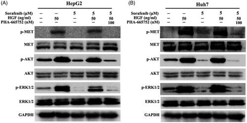
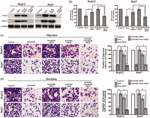
Figure 5 Downregulation of EGR1 by siRNA abrogates the HGF-induced sorafenib resistance. (A) HepG2 and Huh7 cells were transfected with EGR1 siRNA for 48 h, followed by the treatment of HGF (50 ng/ml) for 2 h before cell lysis for Western blot analysis. (B) The transfected HepG2 and Huh7 cells were treated with or without HGF (50 ng/ml) for 24 h, then treated with sorafenib (5 μM) for 72 h. The transfected cells were treated with sorafenib (5 μM) alone or HGF (50 ng/ml) and sorafenib. The cells were then incubated for 24 h (migration assay) or 48 h (invasion assay). (C) Migration abilities were measured using a transwell assay without Matrigel. (D) Invasion abilities were measured using a transwell assay with Matrigel. Data shown represent the mean ± SD. p<.05.